Generic 20 gm betnovate free shippingThe W cells transmit alerts of their optic nerve fibers at a sluggish velocity and receive most of their excitation from rods acne canada scarf betnovate 20 gm on-line, transmitted through small bipolar cells and amacrine cells acne under jawline betnovate 20 gm purchase. They have broad fields within the peripheral retina, are sensitive for detecting directional movement within the field of vision, and are most likely important for crude rod imaginative and prescient under dark circumstances. In addition, because each X cell receives enter from no less than one cone, X cell transmission might be answerable for color vision. The Y cells are the biggest of all and transmit signals to the mind at 50 m/sec or quicker. In primates, a different classification of retinal ganglion cells is used, and as many as 20 kinds of retinal ganglion cells have been described, each responding to a unique characteristic of the visible scene. Some cells respond greatest to particular directions of movement or orientations, whereas others respond to fantastic details, will increase or decreases in light, or specific colours. The two common courses of retinal ganglion cells that have been studied most extensively in primates, together with humans, are designated as magnocellular (M) and parvocellular (P) cells. The P cells (also known as beta cells or, within the central retina, as midget ganglion cells) project to the parvocellular (small cells) layer of the lateral geniculate nucleus of the thalamus. The M cells (also called alpha or parasol cells) project to the magnocellular (large cells) layer of the lateral geniculate nucleus, which, in flip, relays information from the optic tract to the visible cortex, as discussed in Chapter 52. The responses of P cells to stimuli, particularly color stimuli, could be sustained, whereas the responses of M cells are rather more transient. The M cells are rather more sensitive than are P cells to low-contrast, black and white stimuli. The primary capabilities of M and P cells are obvious from their differences: the P cells are highly sensitive to visible indicators that relate to fantastic details and to completely different colours however are relatively insensitive to low-contrast signals, whereas the M cells are highly sensitive to low-contrast stimuli and to rapid movement visual indicators. A third sort of photosensitive retinal ganglion cell has been described that incorporates its personal photopigment, melanopsin. Much much less is known about this cell kind, but these cells seem to ship signals mainly to nonvisual areas of the mind, significantly the suprachiasmatic nucleus of the hypothalamus, the grasp circadian pacemaker. Presumably, these indicators assist management circadian rhythms that synchronize physiological modifications with night time and day. Responses of a ganglion cell to mild in (1) an area excited by a spot of light and (2) an area adjacent to the excited spot. The ganglion cell in this area is inhibited by the mechanism of lateral inhibition. Transmission of Signals Depicting Contrasts within the Visual Scene-The Role of Lateral Inhibition Many ganglion cells respond mainly to distinction borders in the scene, which appears to be the most important means whereby the sample of a scene is transmitted to the mind. When flat gentle is applied to the whole retina, and all of the photoreceptors are stimulated equally by the incident gentle, the distinction sort of ganglion cell is neither stimulated nor inhibited. The cause for this is that indicators transmitted instantly from the photoreceptors by way of depolarizing bipolar cells are excitatory, whereas the alerts transmitted laterally through hyperpolarizing bipolar cells, as well as via horizontal cells, are primarily inhibitory. Thus, the direct excitatory sign via one pathway is more likely to be neutralized by inhibitory alerts via lateral pathways. The two receptors on both sides are related to the same bipolar cell via inhibitory horizontal cells that neutralize the direct excitatory signal if all three receptors are stimulated concurrently by light. Now, allow us to examine what occurs when a distinction border occurs in the visible scene. The incontrovertible fact that one of many lateral photoreceptors is in the dark causes one of many horizontal cells to remain unstimulated. Thus, where visual contrasts happen, the signals through the direct and lateral pathways accentuate each other. In summary, the mechanism of lateral inhibition functions in the eye in the same means that it functions in most different sensory systems-to provide contrast detection and enhancement. It is from the ganglion cells that the long fibers of the optic nerve lead into the brain. Because of the gap involved, the electrotonic method of conduction employed in the rods, cones, and bipolar cells in the retina is now not appropriate; due to this fact, ganglion cells transmit their alerts by the use of repetitive action potentials as an alternative. Furthermore, even when unstimulated, they nonetheless transmit steady impulses at rates varying between 5 and 40 per second. The visual alerts, in turn, are superimposed onto this background ganglion cell firing. The upper panel exhibits rapid impulses for a fraction of a second when a light-weight is first turned on, but these impulses lower rapidly in the subsequent fraction of a second. The decrease tracing is from a ganglion cell situated laterally to the spot of sunshine; this cell is markedly inhibited when the light is turned on because of lateral inhibition. The opposite directions of those responses to gentle are triggered, respectively, by the depolarizing and hyperpolarizing bipolar cells, and the transient nature of the responses might be a minimum of partly generated by the amacrine cells, many of which have similar transient responses themselves. This functionality of the eyes to detect changes in mild depth is strongly developed within the peripheral retina and the central retina. For example, a minute gnat flying across the field of vision is instantaneously detected. Receptor and Neural Function of the Retina Excitation the direct excitatory route by way of a depolarizing bipolar cell, whereas the other shade type inhibits the ganglion cell by the oblique inhibitory route by way of a hyperpolarizing bipolar cell. The significance of these shade distinction mechanisms is that they represent a method whereby the retina begins to differentiate colours. Thus, each color contrast kind of ganglion cell is excited by one color however inhibited by the "opponent" color. Typical association of rods, horizontal cells (H), a bipolar cell (B), and a ganglion cell (G) within the retina, showing excitation on the synapses between the rods and the bipolar cell and horizontal cells however inhibition from the horizontal cells to the bipolar cell. Transmission of Color Signals by the Ganglion Cells A single ganglion cell could additionally be stimulated by a quantity of or only some cones. When all three forms of cones-the red, blue, and green types-stimulate the identical ganglion cell, the signal transmitted by way of the ganglion cell is the same for any shade of the spectrum. Therefore, the signal from the ganglion cell plays no role in the detection of different colors. Conversely, a variety of the ganglion cells are excited by just one shade kind of cone but are inhibited by a second sort. For example, this mechanism incessantly happens for the purple and green cones, with purple causing excitation and green inflicting inhibition, or vice versa. The similar type of reciprocal impact occurs between blue cones on the one hand and a mix of red and green cones (both of that are excited by yellow) then again, giving a reciprocal excitation-inhibition relation between the blue and yellow colors. At the optic chiasm, the optic nerve fibers from the nasal halves of the retinas cross to the alternative sides, where they be part of the fibers from the opposite temporal retinas to form the optic tracts. The fibers of every optic tract then synapse within the dorsal lateral geniculate nucleus of the thalamus and, from there, geniculocalcarine fibers move by way of the optic radiation (also referred to as the geniculocalcarine tract) to the primary visual cortex within the calcarine fissure space of the medial occipital lobe. Thus, the visible pathways can be divided roughly into an old system to the midbrain and base of the forebrain and a new system for direct transmission of visible signals into the visible cortex positioned in the occipital lobes. In people, the brand new system is answerable for notion of virtually all aspects of visual type, colors, and different conscious imaginative and prescient.
20 gm betnovate cheap with mastercardGeneral Principles and Sensory Physiology of -5 mV acne and menopause purchase betnovate 20 gm with visa, which inhibits transmission of the nerve sign through the synapse skin care acne cheap 20 gm betnovate visa. Presynaptic Inhibition In addition to postsynaptic inhibition attributable to inhibitory synapses operating at the neuronal membrane, presynaptic inhibition typically happens on the presynaptic terminals before the signal ever reaches the synapse. Presynaptic inhibition is caused by release of an inhibitory substance onto the outsides of the presynaptic nerve fibrils earlier than their very own endings terminate on the postsynaptic neuron. The unfavorable charges of those ions inhibit synaptic transmission because they cancel a lot of the excitatory effect of the positively charged sodium ions that also enter the terminal fibrils when an motion potential arrives. Presynaptic inhibition happens in many of the sensory pathways within the nervous system. In truth, adjoining sensory nerve fibers typically mutually inhibit each other, which minimizes sideways spread and mixing of signals in sensory tracts. Other types of transmitter substances can excite or inhibit the postsynaptic neuron for for a lot longer periods-for tons of of milliseconds and even for seconds, minutes, or hours. We identified earlier that a change in potential at any single point within the soma will cause the potential to change virtually equally in all places contained in the soma. Therefore, for every excitatory synapse that discharges simultaneously, the total intrasomal potential turns into extra positive by 0. In this final case, the firing threshold had been reached, and an action potential was generated in the axon. This effect of summing simultaneous postsynaptic potentials by activating multiple terminals on extensively spaced areas of the neuronal membrane known as spatial summation. Time Course of Postsynaptic Potentials When an excitatory synapse excites the anterior motor neuron, the neuronal membrane turns into highly permeable to sodium ions for 1 to 2 milliseconds. This potential then slowly declines over the subsequent 15 milliseconds because this is the time required for the surplus constructive costs to leak out of the excited neuron and reestablish the traditional resting membrane potential. However, the changed postsynaptic potential lasts as a lot as 15 milliseconds after the synaptic membrane channels have already closed. Therefore, a second opening of the identical channels can improve the postsynaptic potential to a still higher stage, and the more fast the rate of stimulation, the higher the postsynaptic potential turns into. Facilitation of Neurons Often, the summated postsynaptic potential is excitatory however has not risen excessive sufficient to reach the threshold for firing by the postsynaptic neuron. Consequently, another excitatory sign entering the neuron from another supply can then excite the neuron very simply. The dendrites are long, and their membranes are skinny and a minimum of partially permeable to potassium and chloride ions, making them "leaky" to electrical present. Therefore, earlier than the excitatory potentials can reach the soma, a large share of the potential is misplaced by leakage via the membrane. This decrease in membrane potential because it spreads electrotonically along dendrites towards the soma known as decremental conduction. The farther the excitatory synapse is from the soma of the neuron, the higher will be the decrement and the lesser will be excitatory signal reaching the soma. Therefore, the synapses that lie close to the soma have far more effect in causing neuron excitation or inhibition than people who lie distant from the soma. The dendrites of the anterior motor neurons usually extend 500 to one thousand micrometers in all directions from the neuronal soma, and these dendrites can receive alerts from a big spatial space across the motor neuron. This function supplies an unlimited alternative for summation of indicators from many separate presynaptic nerve fibers. It can be essential that between 80% and 95% of all the presynaptic terminals of the anterior motor neuron terminate on dendrites, in contrast to only 5% to 20% terminating on the neuronal soma. Therefore, a large share of the excitation is provided by alerts transmitted by the use of the dendrites. Most Dendrites Cannot Transmit Action Potentials- But They Can Transmit Signals Within the Same Neuron by Electrotonic Conduction. These inhibitory synapses provide a hyperpolarizing voltage that utterly nullifies the excitatory effect and, certainly, transmits a small quantity of inhibition by electrotonic conduction toward the soma. Transmission of electrotonic current means direct spread of electrical current by ion conduction within the fluids of the dendrites however with out the era of action potentials. Stimulation (or inhibition) of the neuron by this present has particular characteristics, as described subsequent. A highly effective impact of inhibitory synapses on the preliminary section of the axon is also shown. Response characteristics of several sorts of neurons to completely different levels of excitatory state. Also shown within the figure are a quantity of inhibitory synapses positioned instantly on the axon hillock and initial axon phase. This location provides especially powerful inhibition as a end result of it has the direct effect of accelerating the edge for excitation on the very point the place the action potential is normally generated. When the excitatory state of a neuron rises above the edge for excitation, the neuron will hearth repetitively as long as the excitatory state remains at that level. Note that neuron 1 has a low threshold for excitation, whereas neuron 3 has a excessive threshold. But, notice also that neuron 2 has the bottom maximum frequency of discharge, whereas neuron three has the highest maximum frequency. Some neurons within the central nervous system fire continuously as a outcome of even the conventional excitatory state is above the edge stage. Their frequency of firing can usually be increased still more by additional growing their excitatory state. The frequency could be decreased, or firing may even be stopped, by superimposing an inhibitory state on the neuron. Thus, totally different neurons respond in one other way, have completely different thresholds for excitation, and have broadly differing most frequencies of discharge. With somewhat creativeness, one can readily perceive the importance of getting different neurons with these many forms of response traits to perform the widely varying functions of the nervous system. Fatigue is an exceedingly essential characteristic of synaptic perform as a result of when areas of the nervous system turn into overexcited, fatigue causes them to lose this excess excitability after a while. For example, fatigue might be an important means whereby the excess excitability of the mind throughout an epileptic seizure is lastly subdued so that the seizure ceases. Thus, the event of fatigue is a protective mechanism towards excess neuronal activity. This subject is discussed additional in the description of reverberating neuronal circuits in Chapter forty seven. The mechanism of fatigue is especially exhaustion or partial exhaustion of the shops of transmitters within the presynaptic terminals. The excitatory terminals on many neurons can store sufficient excitatory transmitter to trigger only about 10,000 action potentials, and the transmitter may be exhausted in only a few seconds to a few minutes of speedy stimulation. Part of the fatigue course of most likely outcomes from two other components as properly: (1) progressive inactivation of many of the postsynaptic membrane receptors; and (2) gradual improvement of irregular concentrations of ions inside the postsynaptic neuronal cell. Most neurons are extremely responsive to changes in pH of the encircling interstitial fluids.
20 gm betnovate order free shippingThe vasa recta function countercurrent exchangers acne y estres betnovate 20 gm generic with visa, minimizing the washout of solutes from the medullary interstitium acne rosacea treatment betnovate 20 gm order visa. Blood enters and leaves the medulla via the vasa recta at the boundary of the cortex and renal medulla. The vasa recta, like different capillaries, are extremely permeable to solutes within the blood, aside from the plasma proteins. As blood descends into the medulla toward the papillae, it becomes progressively more concentrated, partly by solute entry from the interstitium and partly by loss of water into the interstitium. By the time the blood reaches the ideas of the vasa recta, it has a concentration of about 1200 mOsm/L, the identical as that of the medullary interstitium. As blood ascends back toward the cortex, it becomes progressively less concentrated as solutes diffuse back out into the medullary interstitium and as water moves into the vasa recta. The thick limb of the loop of Henle, distal tubule, and cortical collecting tubule are all less permeable to urea, and solely small quantities of urea reabsorption usually happen in these tubular segments. Under steady-state conditions, the vasa recta carry away solely as a lot solute and water as is absorbed from the medullary tubules, and the Vasa recta mOsm/L 300 Solute 600 Solute 800 Solute 1000 H2O 600 600 350 Solute 600 H2O 800 H2O one thousand 1200 800 Solute 900 one thousand Solute Interstitium mOsm/L 300 excessive focus of solutes established by the countercurrent mechanism is preserved. Certain vasodilators can mark- edly improve renal medullary blood move, thereby washing out a few of the solutes from the renal medulla and decreasing the maximum urine-concentrating capability. Large increases in arterial pressure may also improve the blood flow of the renal medulla to a greater extent than in different areas of the kidney and have a tendency to wash out the hyperosmotic interstitium, thereby reducing urine-concentrating capacity. Plasma flowing down the descending limb of the vasa recta turns into extra hyperosmotic because of diffusion of water out of the blood and diffusion of solutes from the renal interstitial fluid into the blood. In the ascending limb of the vasa recta, solutes diffuse back into the interstitial fluid, and water diffuses back into the vasa recta. Large amounts of solutes can be misplaced from the renal medulla without the U shape of the vasa recta capillaries. However, the proximal tubular membranes are highly permeable to water so, whenever solutes are reabsorbed, water also diffuses by way of the tubular membrane by osmosis. Therefore, the osmolarity of the fluid remains about the identical as the glomerular filtrate-300 mOsm/L. As fluid flows down the de- scending loop of Henle, water is absorbed into the medulla. Thus, the tubular fluid becomes extra dilute because the sodium chloride diffuses out of the tubule and water stays in the tubule. Some of the urea absorbed into the medullary interstitium from the amassing ducts also diffuses into the ascending limb, thereby returning the urea to the tubular system and serving to prevent its washout from the renal medulla. This urea recycling is an additional mechanism that contributes to the hyperosmotic renal medulla. The thick part of the ascending loop of Henle is also just about impermeable to water, but massive quantities of sodium, chloride, potassium, and different ions are actively transported from the tubule into the medullary interstitium. Therefore, fluid in the thick ascending limb of the loop of Henle turns into very dilute, falling to a concentration of about one hundred forty mOsm/L. The early distal tubule has properties much like those of the thick ascending loop of Henle, so additional dilution of the tubular fluid to about one hundred mOsm/L happens as solutes are reabsorbed while water stays within the tubule. Because water reabsorption will increase urea concentration in the tubular fluid, and since the internal medullary amassing ducts have specific urea transporters that tremendously facilitate diffusion, a lot of the highly concentrated urea within the ducts diffuses out of the tubular lumen into the medullary interstitium. This absorption of the urea into the renal medulla contributes to the high osmolarity of the medullary interstitium and excessive concentrating ability of the kidney. First, although sodium chloride is probably considered one of the principal solutes that contribute to the hyperosmolarity of the medullary interstitium, the kidney can, when needed, excrete a extremely concentrated urine that incorporates little sodium chloride. The hyperosmolarity of the urine in these circumstances is due to high concentrations of different solutes, particularly of waste merchandise similar to urea. One condition during which this happens is dehydration accompanied by low sodium intake. Second, massive quantities of dilute urine could be excreted with out rising sodium excretion. Therefore, if massive amounts of solute should be excreted, they must be accompanied by the minimal amount of water essential to excrete them. For example, if 600 milliosmoles of solute should be excreted each day, this requires a minimum of 0. Conversely, when the urine is concentrated, solutes are excreted in extra of water. The complete clearance of solutes from the blood could be expressed because the osmolar clearance (Cosm). This is the volume of plasma cleared of solutes each minute, in the identical way that clearance of a single substance is calculated: Uosm � V Cosm = Posm the place Uosm is the urine osmolarity, V is the urine flow rate, and Posm is plasma osmolarity. For instance, if the plasma osmolarity is 300 mOsm/L, urine osmolarity is 600 mOsm/L, and urine circulate price is 1 ml/min (0. Free Water Clearance-Relative Rates at Which Solutes and Water Are Excreted urine-concentrating ability. When free water clearance is positive, excess water is being excreted by the kidneys; when free water clearance is adverse, excess solutes are being faraway from the blood by the kidneys, and water is being conserved. Using the example discussed earlier, if urine flow price is 1 ml/min and osmolar clearance is 2 ml/min, free water clearance would be �1 ml/min. This implies that as an alternative of water being cleared from the kidneys in excess of solutes, the kidneys are literally returning water to the systemic circulation, as occurs throughout water deficits. Thus, every time urine osmolarity is greater than plasma osmolarity, free water clearance is unfavorable, indicating water conservation. Thus, water free of solutes, called free water, is being lost from the physique, and the plasma is being concentrated when free water clearance is optimistic. The main abnormality observed clinically in folks with this condition is the massive quantity of dilute urine. However, if water consumption is restricted, as can happen in a hospital setting when fluid consumption is restricted or the patient is unconscious. Desmopressin could be given by injection, as a nasal spray, or orally, and it rapidly restores urine output toward normal. In some circumstances, normal or Impairment in the ability of the kidneys to concentrate or dilute the urine appropriately can occur with a quantity of of the next abnormalities: 1. This situation is referred to as nephrogenic diabetes insipidus as a end result of the abnormality resides in the kidneys. In both case, large volumes of dilute urine are shaped, which causes dehydration except fluid consumption is elevated by the identical amount as urine volume is increased. Many types of renal illnesses can impair the concentrating mechanism, especially those who harm the renal medulla (see Chapter 32 for additional discussion). Also, impairment of the operate of the loop of Henle, as happens with diuretics that inhibit electrolyte reabsorption by this section, similar to furosemide, can compromise urineconcentrating capacity.

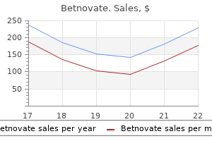
| Comparative prices of Betnovate | | # | Retailer | Average price | | 1 | Raley's | 989 | | 2 | Hy-Vee | 350 | | 3 | Rite Aid | 795 | | 4 | RadioShack | 881 | | 5 | Family Dollar | 875 | | 6 | SonyStyle | 674 | | 7 | Gap | 352 | | 8 | Macy's | 196 | | 9 | IKEA North America | 470 | | 10 | Sports Authority | 882 |
20 gm betnovate mastercardTherefore acne oral medication discount betnovate 20 gm with visa, when reticulocytes leave the bone marrow and pass into the blood stream skin care collagen generic 20 gm betnovate with visa, they continue to form minute portions of hemoglobin for an additional day or so until they become mature erythrocytes. First, succinyl-CoA, which is formed within the Krebs metabolic cycle (as defined in Chapter 68), binds with glycine to type a pyrrole molecule. Each chain has a molecular weight of about 16,000; 4 of these chains, in flip, bind collectively loosely to form the entire hemoglobin molecule. This state of affairs often happens within the illness pernicious anemia, by which the fundamental abnormality is an atrophic gastric mucosa that fails to produce regular gastric secretions. The parietal cells of the gastric glands secrete a glycoprotein referred to as intrinsic issue, which combines with vitamin B12 in meals and makes the B12 available for absorption by the gut in the following means: 1. In this bound state, vitamin B12 is protected from digestion by the gastrointestinal secretions. Still within the sure state, intrinsic factor binds to particular receptor websites on the brush border membranes of the mucosal cells within the ileum. Vitamin B12 is then transported into the blood through the next few hours by the process of pinocytosis, carrying intrinsic issue and the vitamin collectively through the membrane. Basic structure of the heme moiety, showing one of many 4 heme chains that along with globin polypeptide, bind collectively to kind the hemoglobin molecule. The different sorts of chains are designated as alpha chains, beta chains, gamma chains, and delta chains. The commonest form of hemoglobin in adults, hemoglobin A, is a mixture of two alpha chains and two beta chains. Because each hemoglobin chain has a heme prosthetic group containing an atom of iron, and since there are four hemoglobin chains in every hemoglobin molecule, one finds 4 iron atoms in every hemoglobin molecule. Each of those can bind loosely with one molecule of oxygen, making a complete of 4 molecules of oxygen (or eight oxygen atoms) that could be transported by each hemoglobin molecule. The types of hemoglobin chains in the hemoglobin molecule determine the binding affinity of the hemoglobin for oxygen. Abnormalities of the chains can alter the physical traits of the hemoglobin molecule as nicely. For instance, in sickle cell anemia, the amino acid valine is substituted for glutamic acid at one point in every of the two beta chains. These crystals make it virtually impossible for the cells to move through many small capillaries, and the spiked ends of the crystals are more likely to rupture the cell membranes, leading to sickle cell anemia. The and then to launch this oxygen readily within the peripheral tissue capillaries, the place the gaseous tension of oxygen is far lower than within the lungs. Instead, it binds loosely with one of many so-called coordination bonds of the iron atom. The complete amount of iron within the body averages four to 5 grams, about 65% of which is within the form of hemoglobin. About 4% is in the type of myoglobin, 1% is in the form of the assorted heme compounds that promote intracellular oxidation, zero. Transport, storage, and most necessary function of the hemoglobin molecule is its capability to mix loosely and reversibly with oxygen. When iron is absorbed from the small intestine, it immediately combines within the blood plasma with a beta globulin, apotransferrin, to form transferrin, which is then transported in the plasma. The iron is loosely certain in the transferrin and, consequently, can be launched to any tissue cell at any point within the body. Excess iron within the blood is deposited particularly in the liver hepatocytes and less within the reticuloendothelial cells of the bone marrow. Chapter 33 Red Blood Cells, Anemia, and Polycythemia In the cell cytoplasm, iron combines primarily with a protein, apoferritin, to form ferritin. Apoferritin has a molecular weight of about 460,000, and ranging portions of iron can combine in clusters of iron radicals with this massive molecule; due to this fact, ferritin might contain only a small or a large amount of iron. Smaller portions of the iron within the storage pool are in a particularly insoluble kind referred to as hemosiderin. This is very true when the total amount of iron in the body is more than the apoferritin storage pool can accommodate. Hemosiderin collects in cells in the type of large clusters that can be observed microscopically as massive particles. In contrast, ferritin particles are so small and dispersed that they often can be seen within the cell cytoplasm only with an electron microscope. A unique characteristic of the transferrin molecule is that it binds strongly with receptors within the cell membranes of erythroblasts within the bone marrow. There the transferrin delivers the iron on to the mitochondria, where heme is synthesized. There, iron is liberated and is stored primarily within the ferritin pool to be used as wanted for the formation of recent hemoglobin. This slow rate of absorption signifies that even when large quantities of iron are present in the meals, solely small proportions may be absorbed. Conversely, when the iron stores become depleted, the speed of absorption can probably accelerate five or extra times normal. For a lady, additional menstrual lack of blood brings long-term iron loss to a median of about 1. Absorption of Iron From the Intestinal Tract Iron is absorbed from all elements of the small intestine, principally by the following mechanism. The liver secretes moderate amounts of apotransferrin into the bile, which flows by way of the bile duct into the duodenum. Here, the apotransferrin binds with free iron and in addition with certain iron compounds, such as hemoglobin and myoglobin from meat, two of an important sources of iron in the food plan. Then, by pinocytosis, the transferrin molecule, carrying its iron retailer, is absorbed into the epithelial cells and later launched into the blood capillaries beneath these cells within the type of plasma transferrin. The porphyrin portion of the hemoglobin molecule is transformed by the macrophages, via a sequence of levels, into the bile pigment bilirubin, which is launched into the blood and later faraway from the physique by secretion via the liver into the bile. Some forms of anemia and their physiological causes are described in the following sections. For example, exposure to high-dose radiation or chemotherapy for cancer treatment can harm stem cells of the bone marrow, followed in a couple of weeks by anemia. Likewise, high doses of sure poisonous chemical substances, corresponding to insecticides or benzene in gasoline, might trigger the identical effect. In autoimmune issues, such as lupus erythematosus, the immune system begins attacking healthy cells corresponding to bone marrow stem cells, which can lead to aplastic anemia. In about half of aplastic anemia cases the trigger is unknown, a situation known as idiopathic aplastic anemia. Based on the sooner discussions of vitamin B12, folic acid, and intrinsic issue from the abdomen mucosa, one can readily perceive that lack of any one of these can lead to gradual copy of erythroblasts within the bone marrow. Thus, atrophy of the abdomen mucosa, as happens in pernicious anemia, or lack of the entire abdomen after surgical complete gastrectomy can result in megaloblastic anemia. These crystals elongate the cell and provides it the appearance of a sickle somewhat than a biconcave disc. The precipitated hemoglobin additionally damages the cell membrane, so the cells turn into extremely fragile, leading to critical anemia. These antibodies make the Rh-positive cells fragile, leading to speedy rupture and causing the kid to be born with a serious case of anemia.
Betnovate 20 gm buy genericThus acne y embarazo 20 gm betnovate sale, the elements that decide how quickly a fuel will move by way of the membrane are the next: (1) the thickness of the membrane; (2) the floor area of the membrane; (3) the diffusion coefficient of the fuel within the substance of the membrane; and (4) the partial stress distinction of the gas between the two sides of the membrane skin care regimen betnovate 20 gm cheap otc. The thickness of the respiratory membrane occasionally increases-for example, as a end result of edema fluid within the interstitial space of the membrane and within the alveoli-so the respiratory gases should then diffuse not solely by way of the membrane but additionally through this fluid. Also, some pulmonary ailments trigger fibrosis of the lungs, which may improve the thickness of some parts of the respiratory membrane. Because the speed of diffusion through the membrane is inversely proportional to the thickness of the membrane, any issue that will increase the thickness to greater than two to thrice normal can interfere considerably with normal respiratory trade of gases. The surface space of the respiratory membrane may be tremendously decreased by many conditions. For instance, removing of a complete lung decreases the whole floor area to half-normal. Also, in emphysema, many of the alveoli coalesce, with dissolution of many alveolar partitions. Therefore, the new alveolar chambers are much larger than the original alveoli, however the complete surface space of the respiratory membrane is commonly decreased as much as fivefold because of loss of the alveolar walls. Furthermore, gas trade between the alveolar air and pulmonary blood happens via the membranes of all the terminal portions of the lungs, not merely in the alveoli. All these membranes are collectively known as the respiratory membrane, also called the pulmonary membrane. A layer of fluid containing surfactant that lines the alveolus and reduces the floor rigidity of alveolar fluid 2. A thin interstitial area between the alveolar epithelium and capillary membrane 516 Chapter 40 Principles of Gas Exchange; Diffusion of Oxygen and Carbon Dioxide Through the Respiratory Membrane considerably impeded, even under resting situations, and during aggressive sports activities and different strenuous exercise, even the slightest decrease in floor area of the lungs could be a severe detriment to respiratory change of gases. The fee of diffusion within the respiratory membrane is nearly exactly the identical as that in water, for causes explained earlier. The strain difference throughout the respiratory membrane is the distinction between the partial strain of the gas within the alveoli and the partial stress of the gas in the pulmonary capillary blood. Therefore, the distinction between these two pressures is a measure of the online tendency for the fuel molecules to move via the membrane. When the partial stress of a gasoline within the alveoli is greater than the pressure of the fuel in the blood, as is true for O2, internet diffusion from the alveoli into the blood happens. Diffusing capacities for carbon monoxide, oxygen, and carbon dioxide in the regular lungs under resting circumstances and through exercise. All the factors discussed earlier that affect diffusion by way of the respiratory membrane can affect this diffusing capability. In the typical young blood, referred to as the ventilation-perfusion ratio, explained later on this chapter. Therefore, throughout train, oxygenation of the blood is elevated not solely by increased alveolar air flow but in addition by greater diffusing capability of the respiratory membrane for transporting O2 into the blood. The diffusing man, the diffusing capacity for O2 under resting circumstances averages 21 ml/min per mm Hg. The imply O2 strain difference throughout the respiratory membrane during regular quiet respiration is about eleven mm Hg. Multiplying this stress by the diffusing capability (11 � 21) offers a complete of about 230 ml of oxygen diffusing via the respiratory membrane every minute, which is the identical as the rate at which the resting physique uses O2. During strenuous train or other situations that significantly increase pulmonary blood circulate and alveolar air flow, the diffusing capacity for O2 will increase to about three times the diffusing capability under resting conditions. Nevertheless, measurements of diffusion of other gases have shown that the diffusing capacity varies instantly with the diffusion coefficient of the actual gas. The O2 diffusing capacity can be calculated from measurements of the next: (1) alveolar Po2; (2) Po2 within the pulmonary capillary blood; and (3) the rate of O2 uptake by the blood. In Chapter 41, we describe how the normal venous blood (V) has a Po2 of 40 mm Hg and a Pco2 of forty five mm Hg. Therefore, these are additionally the conventional partial pressures of these two gases in alveoli which have blood move but no air flow. This dialogue made the idea that every one the alveoli are ventilated equally, and that blood flow by way of the alveolar capillaries is the same for each alveolus. However, even usually to some extent, and especially in lots of lung illnesses, some areas of the lungs are properly ventilated however have nearly no blood move, whereas different areas may have glorious blood flow however little or no ventilation. In both of those conditions, gasoline trade through the respiratory membrane is significantly impaired, and the particular person could endure extreme respiratory distress, regardless of regular whole ventilation and normal whole pulmonary blood move, however with the air flow and blood flow going to totally different elements of the lungs. Therefore, as a substitute of the alveolar gases coming to equilibrium with the venous blood, the alveolar air becomes equal to the humidified impressed air. Furthermore, because normal inspired and humidified air has a Po2 of 149 mm Hg and a Pco2 of 0 mm Hg, these will be the partial pressures of those two gases within the alveoli. Thus, underneath regular situations, the alveolar air Po2 averages 104 mm Hg and the Pco2 averages 40 mm Hg. When the physiological dead area is nice, much of the work of air flow is wasted effort as a outcome of a lot of the ventilating air never reaches the blood. In a healthy person in the upright position, each pulmonary capillary blood circulate and alveolar ventilation are considerably much less within the higher part of the lung than within the lower half; however, the decrease of blood move is significantly higher than the lower in ventilation. In this space, a small fraction of the blood fails to turn out to be normally oxygenated, and this represents a physiological shunt. However, throughout train, blood flow to the higher a part of the lung will increase markedly, up to now much less physiological dead space happens, and the effectiveness of gasoline exchange now approaches optimum. Also, some extra blood flows via bronchial vessels quite than by way of alveolar capillaries, usually about 2% of the cardiac output; this, too, is unoxygenated, shunted blood. The total quantitative quantity of shunted blood per minute is called the physiological shunt. This physiological shunt is measured in medical pulmonary function laboratories by analyzing the concentration of O2 in each blended venous blood and arterial blood, together with simultaneous measurement of cardiac output. The higher the physiological shunt, the greater the quantity of blood that fails to be oxygenated as it passes by way of the lungs. The ventilation of the anatomical dead house areas of the respiratory passageways is also wasted. The sum of those two forms of wasted ventilation is called the physiological useless space. Thus, in chronic obstructive lung disease, some areas of the lung exhibit critical physiological shunt, and other areas exhibit severe physiological useless space. Both circumstances tremendously decrease the effectiveness of the lungs as gas trade organs, typically reducing their effectiveness to as little as one-tenth normal. In truth, this condition is probably the most prevalent cause of pulmonary disability right now. The presence of hemoglobin in the pink blood cells allows the blood to transport 30 to a hundred times as much O2 as could probably be transported in the form of dissolved O2 in the water of the blood.
Betnovate 20 gm buy low costThus acne en la espalda betnovate 20 gm on-line, when the kidneys are functioning normally skin care education 20 gm betnovate order amex, the chronic renal output curve is much steeper than the acute curve. The highly effective effects of persistent increases in arterial strain on urine output happen because increased strain not only has direct hemodynamic results on the kidney to enhance excretion, but also has oblique results mediated by nervous and hormonal adjustments that happen when blood pressure is increased. Reduced exercise of those antinatriuretic systems due to this fact amplifies the effectiveness of stress natriuresis and diuresis in elevating salt and water excretion throughout chronic will increase in arterial pressure (see Chapters 28 and 30 for additional discussion). Conversely, when blood stress is decreased, the sympathetic nervous system is activated, and formation of antinatriuretic hormones is elevated, including to the direct effects of reduced pressure to decrease renal output of salt and water. This mixture of direct effects of pressure on the kidneys and oblique results of strain on the sympathetic nervous system and varied hormone systems make stress natriuresis and diuresis extremely highly effective factors for long-term management of arterial stress and physique fluid volumes. The importance of neural and hormonal influences on pressure natriuresis is very evident throughout chronic changes in sodium intake. Note that the blood stress equilibrium point B on the curve is almost the identical as level A, the equilibrium level at regular salt intake. Conversely, decreases in salt and water intake to as low as one-sixth normal usually have little impact on arterial stress. In these instances, even reasonable increases in salt consumption could cause vital increases in arterial strain. For example, surgical discount of kidney mass or harm to the kidney as a outcome of hypertension, diabetes, or varied kidney illnesses all trigger blood pressure to be more sensitive to changes in salt consumption. In these instances, greater than normal increases in arterial stress are required to elevate renal output sufficiently to keep a steadiness between the intake and output of salt and water. There is evidence that long-term high salt intake, lasting for a quantity of years, may actually damage the kidneys and eventually makes blood strain extra salt-sensitive. We will discuss salt sensitivity of blood stress in patients with hypertension later in this chapter. Relationships of whole peripheral resistance to the longterm levels of arterial pressure and cardiac output in several clinical abnormalities. Note that altering the whole-body complete peripheral resistance brought on equal and reverse adjustments in cardiac output however, in all circumstances, had no impact on arterial strain. Indeed, when the total peripheral resistance is acutely elevated, the arterial strain does rise instantly. Instead, the arterial pressure returns all the way to regular within about 1 or 2 days. Instead, the kidneys instantly begin to respond to the high arterial pressure, causing stress diuresis and strain natriuresis. Within hours, massive 232 quantities of salt and water are lost from the body; this process continues till the arterial stress returns to the equilibrium pressure degree. At this level, blood stress is normalized, and extracellular fluid volume and blood quantity are decreased to levels beneath normal. Note in all these totally different medical conditions that the arterial strain can also be regular. We will see an example of this mechanism later on this chapter when we talk about hypertension caused by vasoconstrictor mechanisms. The Hypothyroidism Chapter 19 Role of the Kidneys in Long-Term Control of Arterial Pressure and in Hypertension - Increased extracellular fluid quantity Increased blood quantity Increased imply circulatory filling strain Increased venous return of blood to the guts Finally, because arterial stress is the same as cardiac output times complete peripheral resistance, the secondary increase in total peripheral resistance that results from the autoregulation mechanism helps enhance the arterial stress. For example, solely a 5% to 10% increase in cardiac output can improve the arterial pressure from the conventional mean arterial pressure of one hundred mm Hg as a lot as 150 mm Hg when accompanied by an increase in total peripheral resistance because of tissue blood move autoregulation or other elements that trigger vasoconstriction. As salt accumulates in the body, it additionally indirectly increases the extracellular fluid volume for 2 fundamental causes: 1. Although some additional sodium may be saved within the tissues when salt accumulates in the physique, excess salt within the extracellular fluid increases the fluid osmolality. The elevated osmolarity stimulates the thirst center in the mind, making the person drink extra amounts of water to return the extracellular salt concentration to normal and growing the extracellular fluid quantity. The enhance in osmolality caused by the excess salt in the extracellular fluid also stimulates the hypothalamic�posterior pituitary gland secretory mechanism to secrete elevated portions of antidiuretic hormone (discussed in Chapter 29). The antidiuretic hormone then causes the kidneys to reabsorb greatly increased quantities of water from the renal tubular fluid, thereby diminishing the excreted volume of urine but rising the extracellular fluid quantity. Thus, the quantity of salt that accumulates in the body is a crucial determinant of the extracellular fluid volume. Relatively small increases in extracellular fluid and blood volume can often increase the arterial stress substantially. Sequential steps whereby increased extracellular fluid volume will increase the arterial pressure. Note especially that increased cardiac output has both a direct effect to improve arterial stress and an indirect impact by first rising the entire peripheral resistance. The increased arterial strain, in turn, will increase the renal excretion of salt and water and will return extracellular fluid volume to nearly regular if kidney function is regular and vascular capacity is unaltered. Note particularly on this case the two methods during which an increase in cardiac output can improve the arterial stress. One of these is the direct effect of elevated cardiac output to enhance the strain, and the opposite is an indirect impact to elevate complete peripheral vascular resistance through autoregulation of blood flow. Referring to Chapter 17, allow us to recall that each time an excess quantity of blood flows via a tissue, the local tissue vasculature constricts and reduces the blood circulate back towards regular. This phenomenon is recognized as autoregulation, which simply means regulation of blood move by the tissue itself. When increased blood quantity raises the cardiac output, blood move tends to improve in all tissues of the physique; if the increased blood flow exceeds the metabolic wants of the tissues, the autoregulation mechanisms constricts blood vessels all over the body, which in turn increases the total peripheral resistance. A mean arterial pressure greater than a hundred and ten mm Hg (normal is ninety mm Hg) is considered to be hypertensive. Excess workload on the heart leads to early coronary heart failure and coronary heart disease, usually inflicting dying as a result of a coronary heart assault. The high stress regularly damages a major blood vessel within the brain, followed by dying of main parts of the brain; this incidence is a cerebral infarct. Depending on which a part of the brain is involved, a stroke may be deadly or cause paralysis, dementia, blindness, or a number of different critical mind issues. High stress almost all the time causes damage within the kidneys, producing many areas of renal destruction and, eventually, kidney failure, uremia, and dying. Lessons realized from the type of hypertension called volume-loading hypertension have been essential in understanding the function of the renal�body fluid volume mechanism for arterial stress regulation. Volumeloading hypertension means hypertension brought on by excess accumulation of extracellular fluid in the body, some examples of which follow. Experimental Volume-Loading Hypertension Caused by Reduced Kidney Mass and Increased Salt Intake. At the primary circled point on the curve, the two poles of one of the kidneys have been removed, and at the second circled point, the complete opposite kidney was removed, leaving the animals with solely 30% of their normal renal mass. Note that removal of this quantity of kidney mass increased the arterial stress by an average of only 6 mm Hg. Because salt solution fails to quench the thirst, the dogs drank two to four times the normal quantities of quantity, and within a quantity of days, their average arterial pressure rose to about forty mm Hg above regular. After 2 weeks, the dogs got tap water again as a substitute of salt answer; the pressure returned to regular within 2 days.
20 gm betnovate discount visaThe most essential unmeasured cations include calcium acne studios scarf 20 gm betnovate effective, magnesium acne keloid treatment 20 gm betnovate amex, and potassium, and the most important unmeasured anions are albumin, phosphate, sulfate, and different natural anions. Usually, the unmeasured anions exceed the unmeasured cations, and the anion hole ranges between eight and sixteen mEq/L. Kurtz I: Renal tubular acidosis: H+/base and ammonia transport abnormalities and clinical syndromes. Uduman J, Yee J: Pseudo-renal tubular acidosis: situations mimicking renal tubular acidosis. The plasma anion hole is used mainly in diagnosing completely different causes of metabolic acidosis. If plasma sodium focus is unchanged, the focus of anions (Cl- or an unmeasured anion) will increase to keep electroneutrality. Some examples of metabolic acidosis related to a standard or elevated anion gap are proven in Table 31-4. By calculating the anion hole, one can narrow a number of the potential causes of metabolic acidosis. Bibliography Batlle D, Arruda J: Hyperkalemic forms of renal tubular acidosis: clinical and pathophysiological elements. Most diuretics also increase the urinary excretion of solutes, particularly sodium and chloride. In reality, most diuretics which are used clinically act by decreasing renal tubular sodium reabsorption, which causes natriuresis (increased sodium output), in turn causing diuresis (increased water output). That is, in most cases, increased water excretion happens secondary to inhibition of tubular sodium reabsorption because sodium remaining within the tubules acts osmotically to lower water reabsorption. Because renal tubular reabsorption of many solutes, such as potassium, chloride, magnesium, and calcium, is also influenced secondarily by sodium reabsorption, many diuretics increase the renal excretion of these solutes as well. The most common scientific use of diuretics is to cut back extracellular fluid volume, particularly in ailments associated with edema and in hypertension. As mentioned in Chapter 25, lack of sodium from the body mainly decreases extracellular fluid quantity; therefore, diuretics are often administered in medical conditions during which extracellular fluid volume is expanded. Thus, in the steady state, urine output turns into equal to intake, however solely after reductions in arterial stress and extracellular fluid quantity have occurred, relieving the hypertension or edema that prompted using diuretics in the first place. The many diuretics out there for scientific use have different mechanisms of motion and, subsequently, inhibit tubular reabsorption at totally different websites alongside the renal nephron. The common courses of diuretics, their mechanisms of action, and their tubular sites of action are proven in Table 32-1. The osmotic stress of those solutes then reduces water reabsorption, flushing large amounts of tubular fluid into the urine. Large volumes of urine are additionally formed in certain ailments associated with excess solutes that fail to be reabsorbed from the tubular fluid. For instance, when blood glucose concentration rises to excessive levels in diabetes mellitus, the increased filtered load of glucose into the tubules exceeds their capability to reabsorb glucose. Above a plasma glucose concentration of about 250 mg/dl, little of the additional glucose is reabsorbed by the tubules; as a substitute, the excess glucose remains in the tubules, acts as an osmotic diuretic, and increases urine circulate rate. Therefore, one of many hallmarks of uncontrolled diabetes mellitus is polyuria (frequent urination), which is balanced by a excessive degree of fluid consumption (polydipsia) secondary to dehydration, elevated extracellular fluid osmolarity, and activation of the thirst mechanism. These loop diuretics are among the strongest of the clinically used diuretics. Therefore, loop diuretics impair the power of Diuretic remedy Sodium excretion or sodium intake (mEq/day) 200 Excretion the kidneys to concentrate or dilute the urine. Urinary dilution is impaired as a result of the inhibition of sodium and chloride reabsorption within the loop of Henle causes more of these ions to be excreted, along with elevated water excretion. Urine concentrating ability is impaired as a end result of the renal medullary interstitial fluid focus of these ions, and subsequently renal medullary osmolarity, is decreased. Consequently, reabsorption of fluid from the collecting ducts is decreased, so the maximal concentrating capability of the kidneys can additionally be greatly lowered. In addition, decreased renal medullary interstitial fluid osmolarity reduces reabsorption of water from the descending loop of Henle. Because of those multiple results, 20% to 30% of the glomerular filtrate may be delivered into the urine, inflicting urine output, beneath acute circumstances, to be as great as 25 times regular for at least a couple of minutes. Under favorable situations, these agents might cause a maximum of 5% to 10% of the glomerular filtrate to cross into the urine, which is about the same amount of sodium usually reabsorbed by the distal tubules. The quick increase in sodium excretion is accompanied by a lower in extracellular fluid volume. If sodium intake is held fixed, compensatory mechanisms will ultimately return sodium excretion to equal sodium consumption, thus re-establishing sodium balance. Carbonic anhydrase is particularly ample within the proximal tubule, the first web site of motion of carbonic anhydrase inhibitors. Some carbonic anhydrase can be current in different tubular cells, such as within the intercalated cells of the collecting tubule. For example, in 2018, more than 14% of adults in the United States, or greater than 30 million individuals, were estimated to have continual kidney illness, and lots of extra millions have acute renal damage or much less severe types of kidney dysfunction. The time period acute renal failure is often reserved for severe acute kidney injury, by which the kidneys could abruptly stop working totally or virtually completely, necessitating renal replacement remedy corresponding to dialysis, as discussed later in this chapter. Within these two basic categories, there are many particular kidney diseases that may affect the kidney blood vessels, glomeruli, tubules, renal interstitium, and parts of the urinary tract outside the kidney, including the ureters and bladder. In this articler, we talk about particular physiologic abnormalities that occur in a couple of of the more essential forms of kidney ailments. As a consequence, sodium remains within the tubules and acts as an osmotic diuretic, inflicting increased excretion of water, as well as sodium. Because these medication additionally block the effect of aldosterone to promote potassium secretion in the tubules, they lower the excretion of potassium. Mineralocorticoid receptor antagonists additionally cause movement of potassium from the cells to the extracellular fluid. In some cases, this motion causes extracellular fluid potassium concentration to enhance excessively. For this reason, spironolactone and other mineralocorticoid receptor antagonists are referred to as potassium-sparing diuretics. Many of the opposite diuretics cause lack of potassium within the urine, in contrast to the mineralocorticoid receptor antagonists, which spare the loss of potassium. The commonest causes of obstruction of the urinary tract outside the kidney are kidney stones, attributable to precipitation of calcium, urate, or cystine. This decreased activity reduces the transport of potassium into the cells and ultimately decreases the secretion of potassium into the tubular fluid. For this cause, the sodium channel blockers are additionally potassium-sparing diuretics and reduce the urinary excretion rate of potassium.
Generic 20 gm betnovate with visaThe response to adjustments in temperature explains the extreme degree of heat one feels on first coming into a bath of hot water and the acute degree of chilly felt on going from a heated room to outdoor on a cold day skin care kemayoran 20 gm betnovate cheap fast delivery. A few thermal alerts are additionally relayed to the cerebral somatic sensory cortex from the ventrobasal complex skin care doctors edina 20 gm betnovate discount free shipping. Occasionally, a neuron in cortical somatic sensory area I has been found by microelectrode studies to be immediately responsive to both cold or warm stimuli on a specific area of the pores and skin. In other words, thermal detection in all probability results not from direct bodily results of warmth or chilly on the nerve endings however from chemical stimulation of the endings as modified by temperature. However, when a large skin area is stimulated suddenly, the thermal alerts from the entire area are cumulative. A temporary evaluation of these bodily ideas is presented on this chapter, adopted by discussion of the optics of the attention. Light rays ratio of the two refractive indices of the two clear media; and (2) the diploma of angulation between the interface and the getting into wave entrance. The gentle rays passing via the middle of the lens strike the lens exactly perpendicular to the lens surface and, subsequently, move via the lens with out being refracted. Toward either edge of the lens, nonetheless, the light rays strike a progressively more angulated interface. The outer rays bend increasingly toward the center, which known as convergence of travel by way of air at a velocity of about 300,000 km/sec, however they journey much slower via clear solids and liquids. The refractive index of a transparent substance is the ratio of the speed of sunshine in air to the velocity within the substance. Thus, if gentle travels through a specific sort of glass at a velocity of 200,000 km/sec, the refractive index of this glass is 300,000 divided by 200,000, or 1. Refraction of Light Rays at an Interface Between Two Media With Different Refractive Indices. The only impact that happens is decreased velocity of transmission and shorter wavelength, as shown in the figure by the shorter distances between wave fronts. In this determine, the sunshine rays are leaving air, which has a refractive index of 1. When the beam first strikes the angulated interface, the decrease fringe of the beam enters the glass ahead of the higher edge. The wave entrance within the higher portion of the beam continues to journey at a velocity of 300,000 km/sec, whereas that which entered the glass travels at a velocity of 200,000 km/sec. This distinction in velocity causes the upper portion of the wave entrance to transfer forward of the decrease portion so that the wave front is not vertical but is angulated to the right. Because the direction in which gentle travels is all the time perpendicular to the plane of the wave front, the direction of journey of the sunshine beam bends downward. Light rays coming into a glass surface perpendicular to the sunshine rays (A) and a glass floor angulated to the sunshine rays (B). This figure demonstrates that the distance between waves after they enter the glass is shortened to about two-thirds that in air. Bending of sunshine rays at each floor of a convex spherical lens exhibiting that parallel gentle rays are focused to a focal point. Bending of sunshine rays at each surface of a concave spherical lens displaying that parallel mild rays are diverged. Half the bending occurs when the rays enter the lens, and half occurs because the rays exit from the alternative side. If the lens has precisely the right curvature, parallel light rays passing by way of every a half of the lens might be bent precisely sufficient so that all the rays will move via a single level, known as the focal point. This effect is reverse to the impact within the convex lens, and it causes the peripheral gentle rays to diverge from the light rays that move via the center of the lens. Thus, the concave lens diverges mild rays, however the convex lens converges light rays. Cylindrical Lens Bends Light Rays in Only One Plane- Comparison With Spherical Lenses. Note that the cylindrical lens bends light rays from the 2 sides of the lens but not from the highest or the bottom-that is, bending happens in a single airplane but not the other. Conversely, gentle rays that cross by way of the spherical lens are refracted at all edges of the lens (in both planes) towards the central ray, and all of the rays come to a focus. If the check tube is positioned in a beam of sunlight and a bit of paper is introduced progressively closer to the other side of the tube, a certain distance will be discovered at which the sunshine rays come to a focal line. If such a lens is positioned in a beam of daylight, and a chunk of paper is introduced progressively nearer to the lens, the sunshine rays will impinge on a standard point of interest at an acceptable distance. Concave cylindrical lenses diverge gentle rays in just one airplane in the identical method that convex cylindrical lenses converge gentle rays in one airplane. The vertical cylindrical lens converges the light rays that pass through the two sides of the lens, and the horizontal lens converges the top and bottom rays. In different words, two cylindrical lenses crossed at right angles to one another carry out the same function as one spherical lens of the identical refractive power. Focal Length of a Lens the distance past a convex lens at which parallel rays converge to a common focal point is identified as the focal length of the lens. In different phrases, when rays of light which may be already diverging enter a convex lens, the gap of concentrate on the other side of the lens is farther from the lens than the focal length of the lens for parallel rays. This demonstrates that both parallel rays and diverging rays could be focused at the same distance beyond a lens, provided that the lens adjustments its convexity. Because mild rays move by way of the center of a convex lens without being refracted in either path, the light rays from every point source of light are shown to come to a degree give attention to the alternative side of the lens directly consistent with the point source and the center of the lens. Any object in front of the lens is, in actuality, a mosaic of level sources of light. Some of those factors are very shiny and a few are very weak, and so they differ in colour. Each level supply of sunshine on the item involves a separate point concentrate on the other aspect of the lens according to the lens center. However, this picture is the different way up with respect to the unique object, and the 2 lateral sides of the picture are reversed. B, Two cylindrical convex lenses at proper angles to each other, demonstrating that one lens converges light rays in one airplane, and the other lens converges light rays in the airplane at a proper angle. The two lenses mixed give the identical point focus as that obtained with a single spherical convex lens. The two upper lenses of this determine have the same focal size, however the mild rays coming into the highest lens are parallel, whereas these entering the center lens are diverging. The backside lens has far more refractive power than both of the other two lenses. The refractive energy in diopters of a convex lens is the same as 1 meter divided by its focal length. A lens capable of converging parallel light rays to a focus solely 10 centimeters (0. However, if a concave lens diverges light rays on the identical fee that a 1-diopter convex lens converges them, the concave lens is claimed to have a dioptric strength of -1. Likewise, if the concave lens diverges mild rays as a lot as a +10-diopter lens converges them, this lens is claimed to have a power of -10 diopters.
|
