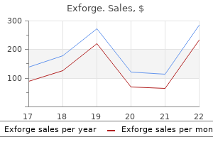
Exforge 80mg cheap otcRuptures within the Descemet membrane may end up in acute corneal hydrops or perhaps a corneal cyst blood pressure simulator purchase 80 mg exforge with mastercard. Corneal topography reveals flattening of the peripheral thinned cornea blood pressure 50 over 0 exforge 80 mg order on-line, with steepening of the corneal surface approximately 90� away from the midpoint of the thinned space. An inflammatory condition of the peripheral cornea which will resemble Terrien marginal degeneration happens in uncommon instances in children and young adults and is also referred to as Fuchs superficial marginal keratitis. This condition might characterize completely different scientific options of the same illness course of. Surgical correction is indicated when perforation is imminent because of progressive thinning, or when marked astigmatism considerably limits vision. Annular lamellar keratoplasty grafts may be required in severe circumstances of 360� marginal degeneration. Peripheral ulcerative keratitis associated with vasculitis manifesting asymmetrically as Fuchs superficial marginal keratitis and Terrien marginal degeneration. Salzmann nodular degeneration Salzmann nodular degeneration is a noninflammatory corneal degeneration that generally occurs as a late sequela to long-standing keratitis such as phlyctenulosis, trachoma, and interstitial keratitis; it could also be idiopathic. The degeneration might not appear until years after the lively keratitis has subsided. They typically develop in a roughly round configuration in the central or paracentral cornea and at the ends of vessels of a pannus. Histologic examination reveals localized replacement of the Bowman layer with hyaline and fibrillar materials, probably representing basement membrane and material much like that found in spheroidal degeneration (discussed earlier). Confocal microscopy reveals elongated basal epithelial cells and activated keratocytes in the anterior stroma, near the nodules; occasionally, subbasal nerves and tortuous stromal nerve bundles are also seen. Treatment for gentle instances is ocular lubrication; manual superficial keratectomy could additionally be indicated in more severe instances (those inflicting decreased vision secondary to irregular astigmatism). A variant of Salzmann nodular degeneration, referred to as peripheral hypertrophic subepithelial corneal degeneration, has been described. Bilateral, pretty symmetric, peripheral, and hypertrophic subepithelial corneal opacification is current. Underlying continual ocular floor irritation is absent, and minimal relief of ocular irritation is achieved with topical corticosteroids. Peripheral hypertrophic subepithelial corneal degeneration: nomenclature, phenotypes, and long-term outcomes. Morphologic and confocal investigation on Salzmann nodular degeneration of the cornea. Corneal keloid Corneal keloids are superficial, typically protuberant, glistening, white corneal masses that can finally involve the entire corneal floor. They are thought to be secondary to a vigorous fibrotic response to corneal injury or continual ocular floor inflammation. Keloids could be congenital or major, they usually have been reported in association with many congenital conditions, such as Lowe disease (oculocerebrorenal syndrome). They have sometimes been confused with hypertrophic scars, Salzmann degeneration, or dermoids. Treatment of symptomatic sufferers might include superficial keratectomy or penetrating or lamellar keratoplasty. Corneal keloid: report of pure history and end result of surgical administration in two instances. Lipid keratopathy In lipid keratopathy, yellow or cream-colored lipids containing ldl cholesterol, neutral fats, and glycoproteins are deposited within the superficial or deeper cornea, usually after prolonged corneal irritation with scarring and corneal vascularization (eg, herpes simplex or herpes zoster keratitis, interstitial keratitis, including syphilitic). Treatment is indicated in instances of decreased imaginative and prescient or compromised beauty look. Lipid keratopathy must be distinguished from Schnyder corneal dystrophy, a uncommon autosomal dominant stromal dystrophy characterised by bilateral corneal opacification resulting from an abnormal accumulation of cholesterol and lipid. Controlling the neovascularization with topical corticosteroids could reduce or even cease progression of the keratopathy. Argon laser therapy with and without fluorescein, photodynamic remedy with verteporfin, and subconjunctival and topical bevacizumab have been reported to cut back corneal neovascularization and lipid deposition. Treatment of herpes zoster�related corneal neovascularization and lipid keratopathy by photodynamic remedy. When the illness is confined to the inner corneal surface, corneal edema may happen because of subnormal endothelial pump operate (Chandler syndrome). The syndrome occurs most commonly in middle-aged ladies and is almost all the time unilateral. They present a attribute reversal of the conventional "light-dark" pattern; thus, the floor seems darkish with an occasional central mild spot, and the intercellular borders seem mild. Varying levels of progressive endothelialization take place in the cornea, in the anterior chamber angle, and on the iris surface. Gonioscopy demonstrates broad-based iridotrabecular synechiae, that are as a result of the proliferation and migration of irregular endothelium over the anterior chamber angle and which end in outflow obstruction and secondary glaucoma. Treatment choices for the corneal part of this syndrome are penetrating keratoplasty and endothelial keratoplasty. Treatment of the glaucoma could be difficult, as filtering surgical procedure could fail due to the progressive growth of the irregular endothelial membrane extending over the trabecular meshwork and filtration website. Implanted tube shunts might overcome regrowth of the membrane within the filtration site. Definitive keratoprosthesis could additionally be an various to repeated conventional keratoplasty. Graft failure and intraocular strain control after keratoplasty in iridocorneal endothelial syndrome. Peripheral cornea guttae Peripheral cornea guttae (Hassall-Henle bodies) are small wartlike excrescences that appear in the peripheral portion of the Descemet membrane. A regular change with growing older, they end result from thickening of the Descemet membrane, which takes place all through life, and happen on the posterior part of the membrane, protruding toward the anterior chamber. With the slit lamp, Hassall-Henle our bodies have the appearance of small, darkish dimples throughout the endothelial mosaic; these are finest seen by specular reflection. Rarely seen before age 20 years, Hassall-Henle our bodies then increase steadily in quantity with age. Central cornea guttae related to progressive stromal and eventually epithelial edema characterize Fuchs endothelial corneal dystrophy (see Chapter 7). Melanin pigmentation Deposits of melanin on the corneal endothelium could be seen in patients with glaucoma associated with pigment dispersion syndrome. The cluster of vertically oriented spindleshaped pigment deposits is normally referred to as Krukenberg spindle (see Table 6-3). These adjustments are accompanied by subconjunctival deposition of fat, which provides the sclera a yellowish appearance.
Diseases - Melioidosis
- Cerebellar hypoplasia
- Scrapie
- Zinc toxicity
- Gordon syndrome
- Axial osteomalacia
- Radio-ulnar synostosis type 2
- Arthrogryposis epileptic seizures migrational brain disorder
- Anophthalmia esophageal atresia cryptorchidism
Order exforge 80mg on-lineIn these sufferers blood pressure pulse rate exforge 80 mg generic free shipping, aspirin significantly lowered the relative odds of a severe vascular occasion by 17% heart attack under 40 80 mg exforge generic visa, comparable to an absolute threat reduction of 30 per 1000 over about 3 years. Benefits of different doses of aspirin Controversy has surrounded probably the most appropriate dose of aspirin, with arguments for daily doses as low as 30 mg to as excessive as 1500 mg [209�211]. First, although not of proven relevance in people, it has been argued that decrease doses ought to inhibit the production of thromboxane A2 by platelets, with out much affecting the production of prostacyclin (a platelet antiaggregant and vasodilator) by endothelial cells, which, unlike platelets, have the biosynthetic equipment to regenerate provides of cyclooxygenase. A single oral dose of a hundred mg is sufficient to nearly completely suppress thromboxane A2 manufacturing in people. Second, because inhibition of cyclooxygenase by aspirin also inhibits the manufacturing of prostaglandins that shield the gastrointestinal mucosa, lower doses of aspirin may produce a smaller extra of bleeding, no less than from the gastrointestinal tract. These doses have been similarly efficient in prevention of significant vascular events [183, 213]. Indirect comparisons of the results of various doses compared with control are much less dependable however the number of outcome events was much greater. Metaanalysis of outcomes for all trials (and 95% confidence interval) is represented by an open diamond. Adjusted management totals have been calculated after converting any unevenly randomized trials to even ones by counting management groups greater than once, but statistical calculations are primarily based on actual numbers from particular person trials. Considering all of the obtainable proof from direct and oblique comparisons in highrisk sufferers, it appears cheap to conclude that aspirin at a dose of 75�150 mg day by day is as efficient as higher doses. Risks of various doses of aspirin A metaanalysis of trials in which participants have been randomized to aspirin or management therapy discovered that aspirin produced a small increased risk of intracranial hemorrhage of about one per a thousand sufferers handled for 3 years. In contrast, observational research have instructed that the danger may be doserelated, but their methodological limitations stop firm conclusions being drawn [217, 218]. Both oblique and direct comparisons found comparable dangers of major extracranial hemorrhage with totally different daily doses [183]. Another metaanalysis discovered the relative excess risk of gastrointestinal bleeding with aspirin to be about 70%, with no definite variation in threat between doses or completely different formulations [219]. Studies that examined the consequences of various doses of aspirin discovered larger dangers for day by day doses >300 mg than for lower doses. The research that reported knowledge on aspirin formulation found similar relative dangers for enteric coated and plain preparations [220]. Randomized trials instantly comparing completely different aspirin doses point out that highdose (500�1500 mg daily) aspirin significantly increases the percentages of higher gastrointestinal symptoms compared with medium dose (75�325 mg daily), and that mediumdose (283 mg daily) is related to a pattern towards an elevated odds of those signs compared with lowdose (30 mg daily) (see Table 17. Aspirin 75�150 mg every day is probably probably the most applicable dose for longterm secondary prevention of significant vascular events to maximize benefits and to decrease opposed effects. This might not necessarily represent a "failure" of remedy, since sufferers who do have additional events whereas on aspirin might need had more or earlier events with out aspirin. Nonplatelet sources of thromboxane A2 (particularly in inflammatory states) or altered thromboxane metabolism. There are numerous potential the cause why sufferers could proceed to have events whereas taking aspirin [222]: Increased turnover of platelets, for instance in an infection, inflammation, or following surgery, could scale back the effectiveness of aspirin. Strategies utilized by some clinicians are to add one other antiplatelet drug, corresponding to modifiedrelease dipyridamole, and even to replace aspirin with an alternate corresponding to clopidogrel (the evidence for various regimens is mentioned in Section 17. Neither is there proof that, for sufferers without atrial fibrillation, oral anticoagulants alone, or together with aspirin, offer any definite net advantage over aspirin alone (Section 17. It is reasonable to consider switching to clopidogrel 75 mg every day or dipyridamole 200 mg twice daily. Poor adherence may be as much of a difficulty with other secondary preventive medication, and this must be discussed with the patient [223]. Genetically decided reduced impact of aspirin: Polymorphisms in the genes encoding cyclooxygenase and other proteins which would possibly be involved in platelet pathways can modify the antiplatelet effects of aspirin, and will account for interindividual variations in laboratory measures of aspirin resistance, and presumably in clinically evident aspirin treatment "failures. However, any variations in results on scientific outcomes between two antiplatelet regimens are likely to be small and so to detect such differences reliably, randomized comparisons would wish to embrace very giant numbers. In terms of antagonistic results, the systematic review found no vital distinction between the thienopyridines and aspirin in intracranial or major extracranial hemorrhage. On the other hand, in contrast with aspirin, the thienopyridines have been associated with a lower risk of gastrointestinal upset and hemorrhage. However, ticlopidine approximately doubled and clopidogrel increased by about onethird the dangers of both pores and skin rash and diarrhea in contrast with aspirin. Ticlopidine, but not clopidogrel, was related to an excess of neutropenia compared with aspirin, but there was no significant difference between the thienopyridines and aspirin in the threat of thrombocytopenia [225]. Ticlopidine has been associated with thrombocytopenia in observational studies, and both ticlopidine and clopidogrel have also been associated in postmarketing surveillance research with thrombotic thrombocytopenic purpura, although that is very uncommon [227�229]. The few trials which have in contrast the 2 thienopyridines immediately have suggested better security and tolerability with clopidogrel versus ticlopidine [230�232], which makes clopidogrel the thienopyridine of choice on security grounds. Although the thienopyridines had a considerably decrease rate of gastrointestinal upset and hemorrhage in contrast with aspirin within the systematic evaluation, probably the most commonly used dose of aspirin within the trials of 325 mg was greater than the currently recommended daily dose of 75�150 mg, which is related to much less gastrointestinal adverse effects, and should examine more favorably with the thienopyridines [225]. In summary, clopidogrel appears to be slightly more practical than aspirin in preventing an ischemic event and might be as protected as aspirin. The giant quantity needed to deal with to see a distinction in vascular outcomes when evaluating aspirin to clopidogrel suggest that aspirin remains the firstchoice antiplatelet drug for all patients at high vascular threat. For sufferers unable to tolerate aspirin because of higher gastrointestinal signs, the addition of an H2blocker might be safer than changing to clopidogrel. Over 20,000 patients have been randomized to every arm; major outcome was recurrent stroke of any type, and secondary outcome was a composite of stroke, myocardial infarction, or death from a vascular cause. Thirtyeight more hemorrhagic strokes occurred within the aspirin plus extendedrelease dipyridamole group, but the number of fatal or disabling strokes remained related in each teams (4. With regard to security, the aspirin plus extendedrelease dipyridamole group had more main hemorrhagic events compared to clopidogrel (4. Noninferiority of aspirin plus dipyridamole in contrast with clopidogrel was not established primarily based on the standards set forth by the investigators; due to this fact, a superiority evaluation was not performed. Interestingly, in tertiary analyses, the aspirin plus extendedrelease dipyridamole group led to a threat discount in new or worsening cases of coronary heart failure. Therefore, aspirin plus extendedrelease dipyridamole was similar to clopidogrel within the prevention of recurrent stroke with a tendency towards more issues. There was no statistically significant distinction between remedy groups in the fee of serious vascular events, however the threat of reasonable to severe bleeding was increased with combination remedy. There was a suggestion that in a subgroup of patients with previous signs of vascular disease combination treatment would possibly achieve web profit, whereas asymptomatic sufferers with a quantity of risk elements might be harmed [239]. Patients within the medical arm had been treated with aspirin at 325 mg every day with clopidogrel seventy five mg for ninety days along with risk factor administration in complete. One group of patients would receive dual antiplatelet therapy with clopidogrel for 90 days plus aspirin for 21 days subsequently followed by placebo through day ninety. The main efficacy end result was any new stroke within ninety days, and the secondary efficacy outcome was a composite of any stroke, myocardial infarction, or vascular dying.
Exforge 80mg purchase free shippingHowever blood pressure 7843 80 mg exforge with amex, if the issue is more likely to blood pressure dizziness exforge 80mg generic visa be long-standing, definitive surgical remedy to right the publicity is really helpful. Most generally, surgical administration consists of permanent lateral and/or medial tarsorrhaphy (see Chapter 13). Insertion of gold or platinum weights into the upper eyelid is also an efficient, more beauty approach to promote eyelid closure. Reported issues of gold weight implants embody an infection, implant malposition, extrusion, induced astigmatism, unacceptable ptosis, and noninfectious inflammatory response to the gold. In addition, correction of any associated eyelid abnormalities, similar to ectropion, entropion, and/or trichiasis, is indicated. For example, in instances of paralytic ectropion of the lower eyelid, a horizontal tightening procedure can also be useful. Neurotrophic Keratopathy and Persistent Corneal Epithelial Defects There are numerous causes of neurotrophic keratopathy, one being injury to cranial nerve V, which finally ends up in corneal hypoesthesia or anesthesia (Table 4-1). Probably the most typical explanation for neurotrophic keratopathy is herpetic keratitis, which may be associated with persistent or recurrent corneal epithelial defects within the absence of replicating virus or active irritation. A neurotrophic cornea also can occur after penetrating keratoplasty or deep anterior lamellar keratoplasty, in the early postoperative period. Some medications used to deal with ocular surface disease and glaucoma could impair epithelial wound healing, resulting within the formation of persistent corneal epithelial defects. In some situations, pericentral pseudodendritiform lesions and pseudogeographic defects may happen. These scientific findings are often misinterpreted as a worsening of the underlying illness and thus could result in extra frequent dosing of the offending medicine. In sufferers with diabetic retinopathy, persistent epithelial defects typically happen following epithelial debridement in the course of vitreoretinal procedures. Diabetic neuropathy is believed to be a possible cause of neurotrophic keratopathy and nonhealing epithelial defects. Persistent corneal epithelial defects typically happen in the central or paracentral cornea and tend to be located inferiorly or inferonasally because of the protecting effect of the Bell phenomenon on the superior cornea. Left untreated, persistent corneal epithelial defects can progress to vascularization and corneal opacification or corneal thinning and possible perforation. Autologous serum drops (20%), which include progress components and fibronectin, may be useful. In circumstances involving significant dry eye, short-term or everlasting punctal occlusion could be efficient in improving the volume and stability of the tear movie and restoring the ocular floor. Patching, low-water-content bandage lenses (soft, thin, highly oxygen-permeable lenses), or scleral contact lenses with a fluid-filled reservoir may facilitate reepithelialization or improve the keratopathy. Corneal crosslinking carried out early in the midst of stromal ulceration has been reported to be helpful in a small variety of patients. Amniotic membrane transplantation has been reported to encourage therapeutic of persistent epithelial erosions. Lateral and/or medial tarsorrhaphy could additionally be required to forestall ocular surface desiccation. Tarsorrhaphy, which may be performed in a temporary or everlasting method, decreases the ocular floor area and tear movie evaporation. Partial or whole conjunctival flaps forestall corneal melting, but they want to be used as a last resort. Floppy Eyelid Syndrome Floppy eyelid syndrome is an ophthalmic disorder consisting of continual ocular irritation and inflammation and characterised by a lax higher tarsus that everts with minimal upward force utilized to the upper eyelid. The syndrome is most incessantly seen in overweight individuals, who usually have obstructive sleep apnea. This may end in trauma to the upper tarsal conjunctiva, inducing inflammation and continual irritation. The condition is most frequently bilateral but could be uneven or unilateral if the affected person always sleeps on the identical facet. Common symptoms embody redness, foreign-body sensation, and mucous discharge most prominent upon waking. The differential analysis contains vernal conjunctivitis, big papillary conjunctivitis, atopic keratoconjunctivitis, bacterial conjunctivitis, and toxic keratopathy. Treatment consists of covering the affected eye with a metallic defend, taping the eyelids closed at night, or performing a surgical process to tighten the upper eyelid. Hyperproliferation, acanthosis, lack of goblet cells, and keratinization are seen in histologic sections of the superior bulbar conjunctiva. However, scrapings or impression cytology of the superior bulbar conjunctiva showing attribute options of nuclear pyknosis with "snake nuclei," increased epithelial cytoplasm�nucleus ratio, goblet cell loss, or keratinization may be useful in the analysis of delicate or confusing instances. Medical therapy options embody topical anti-inflammatory agents, topical cyclosporine, autologous serum eyedrops, and large-diameter bandage contact lenses. Surgical choices embrace thermocauterization of the superior bulbar conjunctiva, resection of the superior limbal bulbar conjunctiva, amniotic membrane transplantation, and conjunctival fixation sutures. Topical cyclosporine A in the treatment of superior limbic keratoconjunctivitis: a long-term follow-up. Development of superior limbic keratoconjunctivitis after upper eyelid blepharoplasty surgical procedure: assist for the mechanical concept of its pathogenesis. Treatment of superior limbic keratoconjunctivitis by thermocauterization of the superior bulbar conjunctiva. Conjunctival fixation sutures for refractory superior limbic keratoconjunctivitis. Conjunctivochalasis Conjunctivochalasis is a time period used to describe laxity or redundancy of the in any other case normal conjunctiva. The tissue could roll up on the eyelid margin or over the lacrimal punctum, in which case it could obstruct tear outflow. Histologic research have revealed elastosis and continual nongranulomatous inflammation. In addition to the conjunctival folds on the eyelid margin, punctate staining may be noticed. This floor disruption is presumably attributable to conjunctival tissue chafing against itself with motion of the attention. A grading system has been proposed (see Meller and Tseng reference) that will assist characterize the findings in this underrecognized condition. Alternatively, the clinician could consider excision of excess conjunctival tissue with main closure to relieve the continual ocular irritation and, in some circumstances, the epiphora. Amniotic tissue grafting and conjunctival fixation are alternative surgical procedures. Correlation between recurrent subconjunctival hemorrhages and conjunctivochalasis by medical profile and profitable surgical consequence. Recurrent Corneal Erosion Recurrent erosions usually happen both in eyes which have suffered a sudden, sharp, abrading corneal harm (eg, from a fingernail, paper reduce, tree branch), in patients with a historical past of herpetic keratitis, or in those with preexisting epithelial basement membrane dystrophy or different dystrophy involving the epithelial basement membrane advanced. The superficial harm produces an epithelial abrasion that heals quickly, regularly leaving no clinical evidence of injury.
Cheap exforge 80 mg overnight deliveryOf the 50% who survive the initial weeks after the hemorrhage arteria inflamada del corazon exforge 80 mg cheap fast delivery, around half are left with everlasting deficits blood pressure medication makes me tired exforge 80mg purchase on-line, usually in highlevel (neuropsychological) functioning, resulting in impairment in their social role [3]. Patients should be transferred as shortly as attainable to a center where a multidisciplinary staff is on the market 24 hours a day, seven days every week, and where enough sufferers are managed to give teams sufficient experience to preserve and improve requirements of care [9]. Patients with apnea or cardiac arrest would require intubation and appropriate cardiovascular resuscitation. A full panel of blood work together with arterial blood gasoline, electrolytes, blood rely, and coagulation parameters must be carried out, and any coagualopathy or thrombocytopenia must be corrected. A routine toxicology display screen must be obtained to examine for medicine corresponding to cocaine. Higher scores on every scale correlate with worse outcomes, with grades of four or 5 typically signifying a considerably greater risk of death or poor outcome [16, 17]. This might lead to structural harm to the myocardium which can be apparent from echocardiography [22], raised troponin I ranges [23], and histological options at postmortem examination together with contraction bands, focal myocardial necrosis, and subendocardial ischemia [24]. Improvement is typical, with no less than some recovery of left ventricle function and improvement in ejection fraction within 1�2 weeks [26, 27]. In acute neurogenic cardiac dysfunction refractory to inotropic or vasopressor administration, a metabolic intervention of induced hyperinsulinemic euglycemia has also been anecdotally reported to be of benefit [31]. Life threatening arrhythmias similar to ventricular fibrillation and "torsades de pointes" could happen [33], however in underneath 5% of sufferers [19, 34]. Pulmonary issues positive endexpiratory pressure ventilation will increase intracranial pressure. Abnormalities warrant additional evaluation with echocardiography to consider cardiac function. Pulmonary edema may result from cardiac dysfunction, or be neurogenic in etiology. Ventilatory assist, optimistic endexpiratory pressure as tolerated by the intracranial stress, and judicious fluid administration should be used to preserve oxygenation. In distinction, current follow is to secure ruptured aneurysms inside 48 hours of bleeding and the emphasis for blood stress management is to stop rebleeding of a ruptured, unsecured aneurysm within the window prior to definitive remedy and the vasospasm risk interval. Furthermore, raised intracranial strain can lead to a large sympathetic discharge, mediated by the anterior hypothalamus, leading to "neurogenic" pulmonary edema. This is thought to lead to elevated permeability of endothelial cells in the pulmonary capillary mattress, resulting in a primarily neurogenic etiology for the edema. The typical scientific image consists of sudden dyspnea, cyanosis, and the manufacturing of pink and frothy sputum. A chest Xray often demonstrates impressive pulmonary edema which may disappear in a matter of hours following constructive endexpiratory pressure ventilation. The management problem is that liberal administration of fluids is usually beneficial for mind perfusion however might delay recovery of pulmonary edema and therefore impair mind oxygenation, and that 15. To guide blood pressure management an arterial line must be placed, particularly for these patients requiring intravenous therapies of elevated blood strain. In sufferers with subarachnoid hemorrhage and unsecured ruptured aneurysm, blood pressure must be managed, with a aim of systemic blood pressure <160 mmHg. Titratable intravenous drips with arterial line monitoring must be used to obtain this aim. In general, the risk of recurrent seizures following an preliminary seizure occurring within the context of an acute mind harm is believed to fall rapidly within the first two weeks, and the convention of prescribing antiepileptic medication for six months is perhaps overly cautious. There are considerations that the physiological response occurring during seizures � including hypertension � may improve the danger of rupture of unsecured aneurysms. The use of phenytoin particularly as prophylaxis was discovered to be predictive of poor neurologic and cognitive outcome in a singlecenter retrospective examine [51]. Gradual obtundation within 24�48 hours of hemorrhage, sometimes accompanied by slow pupillary responses to mild and downward deviation of the eyes, is pretty attribute of acute hydrocephalus [56, 57], and a few sufferers lapse into coma from hydrocephalus within a number of hours of the hemorrhage. Acute hydrocephalus with a considerable amount of intraventricular blood is usually an early indication of the chance of a poor scientific outcome. Additional slices (b) present distinguished frontal (arrow) and occipital (curved arrow) horns, in addition to ballooning of the third ventricle. In sufferers with acute hydrocephalus, exterior ventricular drain placement is the preferred technique for cerebrospinal fluid drainage, permitting each release of cerebrospinal fluid and monitoring of intracranial stress. Lumbar drainage could be thought of however should be strictly avoided in patients with associated intraparenchymal hematoma or obstructive intraventricular hemorrhage to keep away from danger of herniation. The traditional intervention is evacuation of the hematoma with simultaneous clipping of any aneurysm 15. Digital subtraction angiography (c) and threedimensional reconstruction (d) exhibits the large right middle cerebral artery bifurcation aneurysm (curved arrow). Patient required emergent craniotomy for evacuation of hematoma and aneurysm clipping. Hemicraniectomy following evacuation of the hematoma, in anticipation of subsequent edema, can be usually advisable [63]. A modification of this method is to proceed to quick occlusion of the aneurysm by endovascular coiling, adopted by evacuation of the hematoma [64], however this is greatest avoided except there shall be minimal delay launched by the preliminary angiography and coiling. In about 2% of patients � normally within the setting of posterior speaking artery aneurysm � an acute subdural hematoma complicates rupture of an intracranial aneurysm. This seems to be more widespread with recurrent aneurysmal rupture than a primary bleed [65]. Acute subdural hematomas secondary to a ruptured aneurysm, as with all such hematomas, may be shortly life threatening due to mass impact and brain shift; prompt evacuation known as for, with readiness to sort out the underlying aneurysmal source surgically at the similar time [67]. Anxiolytic drugs or artificial opiates corresponding to tramadol can also be helpful in acquiring ache aid. Constipation is a standard drawback with all opiates and prophylaxis with stool softeners is important to stop gastrointestinal complications. For as lengthy as the aneurysm has not been occluded the patient ought to be kept on bedrest, with as few exterior stimuli as possible, on the idea that any form of pleasure might affect blood stress and improve the risk of rebleeding. Venous access A dependable intravenous route ought to be maintained for emergency administration of blood strain brokers, plasma expanders, or antiepileptic medicine; central venous entry is most well-liked as it could additionally provide central venous strain monitoring within the postaneurysm remedy interval as properly as administration of medicines, vasoactive agents, fluids, colloids, or hypertonic solutions as essential. Any change in the level of consciousness or new confusion or focal neurological signs may signify rebleeding, hydrocephalus, hypoxia, early cerebral ischemia, seizures, an infection, or a systemic medical complication. Frequent reexamination and workup for treatable causes of altered consciousness or confusion should be ongoing. Condom gadgets leak or slip off too usually to be of practical use in these critically unwell patients. Gastrointestinal management Gastrointestinal prophylaxis in opposition to stress ulcers with H2 blockers ought to be thought of for all patients, and specifically for mechanically ventilated sufferers or these with a previous history of gastroesophageal reflux or ulcers. Straining ought to be avoided if at all attainable because of the attendant surges in arterial blood stress, and stool softeners should be prescribed routinely. Enemas should be prevented as a result of the potential for marked improve in intraabdominal stress and, secondarily, intracranial strain. Antiemetics must be out there, to avoid the intracranial stress surges related to emesis. To scale back threat of rebleed and intracranial pressure elevation, patients with unsecured ruptured aneurysms should be managed on bedrest, with analgesics, antiemetics, and stool softeners, as nicely as stomach acid prophylaxis and bladder catheterization.
Eurphrasia rostkoviana (Eyebright). Exforge. - Dosing considerations for Eyebright.
- Use directly on the eye for eye conditions, including fatigue, inflammation, infections, and other conditions.
- What is Eyebright?
- Inflamed nasal passages, inflamed sinuses (sinusitis), colds, allergies, coughs, earaches, headache, and many other uses.
- How does Eyebright work?
- Are there safety concerns?
Source: http://www.rxlist.com/script/main/art.asp?articlekey=96151
Exforge 80mg order fast deliveryFurthermore arrhythmia journal cheap 80 mg exforge free shipping, this research was revealed previous to heart attack 8 trailer 80 mg exforge with amex our present understanding of pelvic parameters. The influence of pelvic tilt on the deformities as well as achieving a balanced relationship between the pelvic incidence and the lumbar lordosis postoperatively, which are known key determinants of medical outcome,2 have been due to this fact not addressed in this series. Sagittal steadiness refers to whether the center of mass is centered over the pelvis and ft. Chapter forty six � Pedicle Subtraction Osteotomy 235 the selection of osteotomy must be based mostly on the morphology and rigidity of the deformity. This contributed to a really low pseudarthrosis rate at the level of the osteotomy of 3. Current studies describe techniques that involve higher posterior bone resection and depart gaps in the posterior column following osteotomy closure. Two-rod constructs and adjacent interbody fusion may reduce the rate of early rod fracture. Reducing these structures to one another with osteotomy closure achieves direct bone apposition, restoring the integrity of the posterior column. This research had a big influence in demonstrating the results of pedicle subtraction osteotomies in addressing extreme sagittal malalignment. With better understanding of sagittal parameters and the limitations and strengths of assorted osteotomies, refining of the indications and surgical strategies will additional define the function of pedicle subtraction osteotomies within the remedy of mounted spinal deformities. Pedicle subtraction osteotomy for the treatment of fastened sagittal imbalance: Surgical technique. Classification of sagittal imbalance based on spinal alignment and compensatory mechanisms. An analysis of sagittal spinal alignment in one hundred asymptomatic middle and older aged volunteers. Successful correction of sagittal imbalance may be calculated on the premise of pelvic incidence and age. Comparison of Smith-Petersen versus pedicle subtraction osteotomy for the correction of fixed sagittal imbalance. Comparison of pedicle subtraction and Smith-Petersen osteotomies in correcting thoracic kyphosis when closed with a central hook-rod construct. Assessment of symptomatic rod fracture after posterior instrumented fusion for grownup spinal deformity. Reducing rod breakage and pseudarthrosis in pedicle subtraction osteotomy: the importance of rod number and configuration in 264 patients with 2-year follow-up. Construct rigidity after fatigue loading in pedicle subtraction osteotomy with or without adjoining interbody structural cages. Neurosurgery 48(3):569�574, 2001 Reviewed by James Stenson and Kris Radcliff Research Question/Objective 47 Circumferential fusion improves charges of fusion and clinical outcomes when utilized to deal with lumbosacral backbone pathologies. Historically, surgeons had the selection of both a sequential two-step anterior and posterior fusion or posterior lumbar interbody fusion with simultaneous posterolateral fusion to obtain circumferential fusion. However, these approaches are fraught with their respective individual complications. The authors tried to study and report their findings of ache aid after surgical procedure, intraoperative issues, infection price, and postoperative problems. Five patients beforehand underwent lumbosacral backbone surgical procedure, certainly one of which was an tried lumbar fusion. Transforaminal lumbar interbody fusion: Technique, issues, and early results. Intervention Results Low back pain dissipated fully in 16 sufferers following surgical procedure. Five patients had moderately persistent low again ache requiring oral narcotics for ache management. Fortunately, the index procedure resolved radicular symptoms in all 19 sufferers who introduced with radiculopathy at the time of surgery. Additional postoperative complications included transient postoperative brachial neuralgia secondary to intraoperative positioning, distal neuropathy in the arm due to extended blood stress cuff inflation, and L5 motor weak spot. The patient with distal arm neuropathy passed away three months following surgical procedure from cardiomyopathy. The acknowledged affected person was nonetheless affected by distal neuropathy on the time of death. The L5 motor weakness was discovered solely when the affected person tried to resume jogging. Second, the already limited pattern measurement is further hindered by a relatively short follow-up period. Longer-term follow-up can be perfect because it takes as much as 12 months for fusion rates to be decided. Although not supposed to be an outcome study, the article would profit from goal and validated ache and performance scales such as the preoperative and postoperative Visual Analog Scale and the Oswestry Disability Index. Furthermore, affected person choice was not mentioned within the text, which created an inherent bias for the examine pool. The conventional open approach requires a big gentle tissue dissection to expose anatomic landmarks for pedicle screw fixation, leading to increased postoperative pain, longer recovery, and impaired spinal function. Radiographic restoration of lumbar alignment after transforaminal lumbar interbody fusion. Transforaminal lumbar interbody fusion: A retrospective research of long-term ache relief and fusion outcomes. Transforaminal lumbar interbody fusion: Surgical method and ends in 24 sufferers. Transforaminal lumbar interbody fusion: Clinical and radiographic outcomes and complications in one hundred consecutive sufferers. The difference of surgical website infection in accordance with the strategies of lumbar fusion surgical procedure. Comparison of medical and radiological outcomes of posterolateral fusion, posterior lumbar interbody fusion and transforaminal lumbar interbody fusion methods in the treatment of degenerative lumbar backbone. Analytical comparison research of the clinical and radiological consequence of backbone fixation utilizing posterolateral, posterior lumber interbody and transforaminal lumber interbody spinal fixation techniques to deal with lumber backbone degenerative disc disease. The cost-effectiveness of interbody fusions versus posterolateral fusions in 137 patients with lumbar spondylolisthesis. A randomized managed trial comparing transforaminal lumbar interbody fusion and uninstrumented posterolateral fusion in the degenerative lumbar spine. Transforaminal lumbar interbody fusion in degenerative disk disease and spondylolisthesis Grade I: Minimally invasive versus open surgical procedure. Transforaminal lumbar interbody graft placement using an articulating supply arm facilitates increased segmental lordosis with superior anterior and midline graft placement. Kiehna forty eight Research Question/Objective Spinal twine accidents in youngsters are extremely uncommon; nevertheless, acute diagnosis in this inhabitants is difficult. The juvenile spine has inherent elasticity that allows for self-reduction of intersegmental injuries, which leads to a standard showing scientific radiograph. Follow-Up Biweekly by way of the interval by which they wore a brace, then for 1 12 months past the damage. All sufferers acquired complete cervical spine movies including open-mouth odontoid views.
Generic exforge 80mg overnight deliveryWithout an iridotomy hypertension and kidney disease exforge 80mg purchase amex, closure of the angle normally progresses and makes the subsequent glaucoma harder to control prehypertension 2016 generic 80 mg exforge otc. Even with a patent peripheral iridotomy, progressive angle closure can occur, and repeated periodic gonioscopy is crucial. Angle closure in plateau iris is most frequently brought on by anteriorly positioned ciliary processes that critically slim the anterior chamber recess by pushing the peripheral iris forward. Evidence means that plateau iris configuration may result from a extra anterior junction of the iris dilator muscle and the ciliary epithelium, which causes the iris root to be more articulated. The angle may be additional compromised following dilation of the pupil because the peripheral iris bunches up and obstructs the trabecular meshwork. Plateau iris could additionally be suspected if the central anterior chamber appears to be of regular depth and the iris plane seems flat for an eye with angle closure. The situation might be missed if the examiner relies solely on the slit-lamp examination or the Van Herick methodology of angle examination. Note that the midperipheral angle appears deeper (double arrow) than the slim angles related to pupillary block. B, Plateau iris syndrome following laser peripheral iridoplasty, with a a lot deeper angle recess (arrow). Eyes with plateau iris syndrome stay predisposed to angle closure despite a patent iridotomy, because of the peripheral iris anatomy. Thus, cautious assessment of the angle following iridotomy or lensectomy is critical to decide whether further treatment to additional deepen the angle is required. Repeated gonioscopy at regular intervals is important because of the chance of persistent angle closure regardless of measures to deepen the angle recess. Prevalence and traits of plateau iris configuration amongst American Caucasian, American Chinese and mainland Chinese subjects. Plateau iris syndrome: changes in angle opening related to darkish, mild, and pilocarpine administration. Secondary Angle Closure With Pupillary Block Lens-Induced Angle Closure Phacomorphic glaucoma the mechanism of phacomorphic glaucoma is usually multifactorial. However, by definition, a significant factor of the pathologic angle narrowing is related to the acquired mass impact of the cataractous lens itself. Phacomorphic narrowing of the angle usually occurs slowly with formation of the cataract. In many circumstances, the iridotomy is pointless, as cataract surgery is the definitive therapy in eyes that have the potential for improved imaginative and prescient. Cholinergic brokers have a minimal function within the remedy of this situation as a result of they might further slender the angle and worsen the vision in the presence of cataract. Lens intumescence precipitates pupillary block and secondary angle closure in an eye not anatomically predisposed to angle closure. C, In this case, a extra long-term answer is offered by thinning the peripheral iris with argon laser iridoplasty. With ahead displacement, pupillary block may occur, leading to iris bomb�, shallowing of the anterior chamber angle, and secondary angle closure. Lens extraction is usually indicated to restore imaginative and prescient and to reduce the risk of recurrent pupillary block and persistent angle closure development. Treatment with cycloplegia might tighten the zonule, flatten the lens, and pull it posteriorly, breaking the pupillary block. Miotics may make the condition worse by rising the pupillary block and by rotating the ciliary body ahead, loosening the zonule and allowing the lens to become more globular. Microspherophakia is usually familial and may happen as an isolated situation or as part of both Weill-Marchesani or Marfan syndrome. Aphakic or pseudophakic angle-closure glaucoma Pupillary block may happen in aphakic and pseudophakic eyes. An intact vitreous face can block the pupil and/or an iridotomy website in aphakic or pseudophakic eyes or in a phakic eye with a dislocated lens. Generally, the anterior chamber shallows and the iris shows appreciable bomb� configuration. Treatment with mydriatic and cycloplegic agents may restore the aqueous flow by way of the pupil however may also make performing a laser iridotomy troublesome initially. The lens (arrow) is trapped anteriorly by the pupil, resulting in iris bomb� and a dramatic shallowing of the anterior chamber. B, Gonioscopic view of the identical eye reveals that the dislocated lens is within the inferior vitreous cavity. Pupillary block develops with apposition of the iris, vitreous face, and/or lens optic. The lens haptic or vitreous might hinder the iridectomy site or the pupil, and the peripheral iris bows ahead across the anterior chamber intraocular lens to occlude the angle. The central chamber remains deep relative to the peripheral chamber on this instance, because the 132 Glaucoma lens haptic and optic prevent the central portions of the iris and vitreous face from shifting ahead. In addition, pupillary block might happen following posterior capsulotomy when vitreous obstructs the pupil. A situation referred to as capsular block may be seen, whereby retained viscoelastic or fluid within the capsular bag pushes a posterior chamber intraocular lens anteriorly, which can slender the angle. Secondary Angle Closure Without Pupillary Block A variety of disorders can result in secondary angle closure with out pupillary block. The illness is characterized by fantastic arborizing blood vessels on the floor of the iris, pupillary margin, and trabecular meshwork, which are accompanied by a fibrous membrane. Table 5-4 Disorders Predisposing to Neovascularization of the Iris and Angle Systemic vascular disease Carotid occlusive disease* Carotid artery ligation Carotid-cavernous fistula Giant cell arteritis Takayasu (pulseless) disease Ocular vascular illness Diabetic retinopathy* Central retinal vein occlusion* Central retinal artery occlusion Branch retinal vein occlusion Sickle cell retinopathy Coats disease Eales illness Retinopathy of prematurity Persistent fetal vasculature Syphilitic vasculitis Anterior phase ischemia * Most widespread causes. While performing gonioscopy in sufferers with attainable neovascularization, the clinician might discover it helpful to use a shiny slit-lamp beam of sunshine and high magnification in order to greatest visualize these nice vessels. In uncommon situations, anterior segment neovascularization might occur without demonstrable retinal ischemia, as in Fuchs heterochromic uveitis and different forms of uveitis, pseudoexfoliation syndrome, or isolated iris melanomas. In establishing a correct prognosis, the clinician ought to distinguish dilated iris vessels related to irritation from newly formed abnormal blood vessels. Because the prognosis for neovascular glaucoma is poor, prevention and early diagnosis are important. Since the most common cause of iris neovascularization is ischemic retinopathy, the definitive therapy when the ocular media are clear is panretinal photocoagulation. Even in the presence of complete synechial angle closure, panretinal photocoagulation may enhance the success fee of subsequent glaucoma surgery by eliminating the angiogenic stimulus and should decrease the danger of hemorrhage on the time of surgery. Medical management of neovascular glaucoma yields variable success and is only a temporizing measure until more definitive incisional or laser surgery is undertaken. If these therapies fail or if the attention has poor visible potential, both endoscopic or transscleral cyclophotocoagulation can be thought-about as a substitute for filtering surgical procedure or tube shunt implantation.
Purchase 80mg exforge overnight deliverySample Size Three hundred fifty-eight sufferers had been assessed for eligibility blood pressure food exforge 80mg overnight delivery, and 247 patients underwent randomization hypertension nursing teaching 80 mg exforge cheap otc. One hundred twenty-three patients, including 67 with degenerative spondylolisthesis, were assigned to the decompression-with-fusion group, and 124 patients, including 68 with degenerative spondylolisthesis, were assigned to the decompression-alone group. Follow-Up Surveys had been despatched to participants before surgery and at 1, 2, and 5 years postoperatively. Exclusion criteria have been spondylolysis, degenerative lumbar scoliosis, history of previous surgery for lumbar spinal stenosis or instability, stenosis not brought on by degenerative change, stenosis brought on by herniated disc, other spinal circumstances. Intervention or Treatment Received Patients in the decompression-alone group obtained decompression surgery, whereas patients in the decompressionwith-fusion group obtained decompression with fusion. There were no significant differences between the two groups in any of the preoperative variables. Among patients with preoperative degenerative spondylolisthesis, the mean degree of vertebral slip was 7. There were additionally no significant variations between the 2 teams within the secondary functional, pain, and quality-of-life measures (Table 32. The decompression-with-fusion group had significantly greater operative time and quantity of bleeding. The reoperation rate was 22% within the fusion group and 21% within the decompression-alone group. In phrases of well being financial analysis, the imply direct prices of fusion had been $6800 greater than decompression alone, mostly as a outcome of further working time, extended hospitalization, and price of the implant. The data mirror the number of sufferers who answered "fully gone," "significantly improved," or "considerably improved. Chapter 32 � A Randomized, Controlled Trial of Fusion Surgery 169 Study Limitations First, the inclusion and exclusion criteria allowed into the study a wider, more heterogeneous study inhabitants, including those with lumbar stenosis with or with out degenerative spondylolisthesis. Additionally, the diploma of spondylolisthesis was not properly characterised, other than noting that the average diploma of slip was 7. Given these limitations, drawing conclusions from this research about particular disease populations could additionally be difficult. Second, the type of fusion or decompression surgical procedure was determined solely by the surgeon. The thresholds for reoperation are unclear and also were determined solely by the surgeon. The function of fusion in degenerative spondylolisthesis and spinal stenosis is a hotly debated and controversial topic. In 1991, Herkowitz and Kurz published a potential examine of fifty patients evaluating decompressive laminectomy with decompressive laminectomy and concomitant intertransverse-process arthrodesis, and found significantly better leads to the fusion group with respect to reduction of again and leg ache. Furthermore, they argued that the upper fee of reoperation in the decompression-alone group could offset any well being care costs saved from decompression alone with out fusion. Shortly following this, a Cochrane systematic review, which included the above two research in addition to 22 other randomized control trials for the therapy of lumbar spinal stenosis, concluded that decompression plus fusion has not been proven to be superior to decompression alone. More analysis is required to delineate patient traits which may be predictive of which surgical technique. For the time being, spine surgeons should continue to be cognizant of the concept, while fusion is a more invasive operation with larger complication rates and longer recovery durations, it could be used to forestall the progression of listhesis and instability and to reduce the necessity to reoperate at that degree. National trends in the use of fusion methods to deal with degenerative spondylolisthesis. Is there a task for decompression alone for treating symptomatic degenerative lumbar spondylolisthesis The evidence on surgical interventions for low again disorders: An overview of systematic evaluations. The surgical management of degenerative lumbar spondylolisthesis: A systematic evaluation. Trends, main medical issues, and expenses related to surgical procedure for lumbar spinal stenosis in older adults. The earlier King classification only thought-about the deformity in the coronal plane and acknowledged only 5 curve types. Furthermore, it lacked reliability and reproducibility with poor-to-fair validity. Interobserver and intraobserver reliability was estimated by calculating the kappa coefficient values of easy and weighted components with 95% confidence intervals. A single observer subsequently and retrospectively utilized the brand new Lenke system to consecutively operated patients in order to assess the prevalence of curve sorts. Adolescent idiopathic scoliosis: A new classification to decide extent of spinal arthrodesis. A single observer retrospectively assessed 315 consecutively operatively treated patients for the aim of figuring out the prevalence of curve types. Results A classification system based on three components-curve type, a lumbar modifier, and a sagittal thoracic modifier-was developed. In figuring out the curve kind with the new classification, the mean interobserver reliability was 93% (range, 85%�100%) with a imply kappa value of zero. In reviewing the King classification, the same 5 reviewers revealed a mean interobserver reliability of 64%, with a imply kappa value of 0. The intraobserver reliability of the model new classification was 85% (range, 72%�100%), with a mean kappa worth of 0. For group 2, the kappa values for interobserver reliability with the new classification have been 0. With the exception of curve kind interobserver reliability, these values all symbolize good-to-excellent reliability. In applying the model new classification to the consecutive sequence of 315 patients, sort one (main thoracic) was the most prevalent kind, present in 126 (40%) of the 315. The prevalence of curve kind 4 (triple major) and 6 (thoracolumbar/lumbar-main thoracic) had been each 3%. The prevalence of the lumbar spine modifier types A, B, and C was 30%, 21%, and 49%, respectively. The sagittal thoracic modifier revealed hypokyphosis in 18%, normal kyphosis in 71%, and hyperkyphosis in 11%. Study Limitations An increased factor of complexity exists in creating the new Lenke system. Forty-two curve types could be derived when lumbar Chapter 33 � Adolescent Idiopathic Scoliosis 175 and thoracic sagittal modifiers are utilized to 6 well-established curve types. Despite this, solely 27 instances were assessed in figuring out the interobserver and intraobserver reliability. A bias might exist, nonetheless, among group 1, as developers of the new Lenke system and critics of the King classification.
80mg exforge purchase otcDiagnosis and remedy of pulmonary arteriovenous malformations in hereditary hemorrhagic telangiectasia: an overview hypertension journal impact factor buy exforge 80 mg free shipping. Ruptured intracranial aneurysm associated with von HippelLindau 461 462 463 464 465 466 467 468 syndrome: a molecular link Imaging characteristics of cerebrovascular arteriopathy and stroke in HutchinsonGilford progeria syndrome blood pressure quizzes exforge 80mg buy free shipping. Pathogenesis and radiobiology of mind arteriovenous malformations: implications for risk stratification in pure history and posttreatment course. The presence or absence of different elements that may be of minor significance individually, such as old age, black race, and low cholesterol degree may be decisive [2, 3]. Structural: disorders and malformations of the cerebral blood vessels Chronic hypertension (Section 8. A systematic evaluate of research that in contrast blood stress in deep versus lobar hemorrhage demonstrated moderately higher prestroke extra of hypertension (odds ratio 1. Furthermore, sufferers categorized as normotensive in these research might need had gentle levels of hypertension with out proof of endorgan harm. As in another dysfunction, highrisk sufferers account for only a small proportion of the inhabitants, with most sufferers solely at average danger of creating the disease. Charcot�Bouchard aneurysms and, together with cerebral amyloid angiopathy leading to lobar hemorrhage, accounts for many circumstances in aged people. The proportion of each causative factor inside a given affected person population is determined by the age distribution and presence of different risk components. The relative chances of every consider relation to hemorrhage location are additional discussed in Section eight. Chronic hypertension underlies the degenerative adjustments in the small perforating arteries that ultimately result in their rupture in deep subcortical and infratentorial areas. Hypertension will increase the risk of hemorrhagic and ischemic stroke to an identical diploma [8], doubling the danger with every 5 mmHg rise within the diastolic blood strain [9]. They found multiple, minute outpouchings of small blood vessels that they called miliary aneurysms or microaneurysms [14]. In 1963, Ross Russell carried out a postmortem examine in a gaggle of hypertensive and normotensive aged topics and demonstrated an in depth relationship between microaneurysms and hypertension [17]. Microaneurysms had been present in 15 of 16 individuals with hypertension, whereas solely 10 of 38 normotensive topics had microaneurysms. In addition, the number of microaneurysms per person was considerably greater in the hypertensive group. It is likely that the rupture of the small perforating arteries outcomes from lesions other than microaneurysms. He demonstrated degenerative adjustments including segmental lipohyalinosis and fibrinoid necrosis within the wall of ruptured perforating arteries. These adjustments have been associated with arterial wall thinning, making the arteries vulnerable to microtrauma ensuing from adjoining hemorrhage. Furthermore, arterial dissection was proven to be widespread characteristic in ruptured perforating arteries [22]. Postmortem studies confirmed the presence of hemosiderin in about twothirds of these lesions [25]. Microbleeds are seen in 5�23% within the basic inhabitants, with roughly 75% of lesions located within the cerebral cortex or subcortical white matter, and less regularly in the basal ganglia and inside capsule [26�29]. Barium sulfate was injected into the arterial tree before fixation of the brain with formalin. Along with age and low serum ldl cholesterol level, hypertension is the most common threat factor for microbleeds in healthy individuals as well as these with stroke [36, 37]. Microbleeds are most likely to be more widespread in sufferers taking antiplatelets or anticoagulants [38�40]. Microbleeds are common in patients with stroke [41, 42] and appear to predict the incidence of future ischemic and hemorrhagic stroke in healthy individuals [43] as well as those that have had transient ischemic assault or ischemic stroke [44]. Hemorrhagic transformation of ischemic stroke, whether spontaneous or thrombolysisassociated, tends to be more widespread in sufferers with microbleeds [45]. Approximately 25% of patients demonstrate vital hematoma growth, particularly through the preliminary days after hemorrhage [46]. Significant hematoma enlargement is outlined as 33�50% quantity increase or an absolute improve in hematoma volume of 12. Hematoma expansion is likely a heterogeneous process involving breakdown of blood�brain barrier and dysregulation of hemostasis [48]. Miller Fisher proposed that rupture of a single small artery might lead to subsequent rupture of different fragile arteries inflicting an "avalanche" of secondary hemorrhages [20]. An additional 12% of sufferers had growth between 1 and 24 hours after presentation. Anticoagulation is related to increased risk of early and delayed hematoma expansion, as well as greater mortality [51]. Antiplatelets doubtless increase the chance of hematoma enlargement, though lower than anticoagulants [52]. This may be as a result of the vulnerability of these areas to minor trauma, or characteristics of the posterior circulation that influence elimination of amyloid [71]. Left: Right basal ganglia hemorrhage (white arrow) with rupture into the frontal horn of the proper lateral ventricle (open arrow). Right: Second hemorrhage eight months after the preliminary episode; cerebral angiography was regular. The most frequent situation is cognitive impairment, generally presenting as fullblown dementia with out preceding hemorrhage [77, 78]. It is usually associated with diffuse demyelination of subcortical white matter much like abnormalities seen in patients with hypertensive vasculopathy [82, 83]. However, white matter abnormalities, also identified as leukoaraiosis, are thought to outcome from arteriolosclerotic adjustments within the perforating arteries. These episodes may be attributable to transient ischemia or small hemorrhages that end in focal seizures or spreading cortical despair [86]. Often, the diagnosis could be made solely in retrospect when a lobar hemorrhage supervenes. The presence of microbleeds on susceptibility weighted pictures within the corresponding mind region [87] and the absence of hemodynamically vital stenosis in that vascular territory assist distinguish these spells from transient ischemic assault. A particular analysis can solely be made with mind biopsy or postmortem examination, demonstrating perivascular irritation with multinucleated big cells and amyloidladen vessels. Genetics of sporadic cerebral amyloid angiopathy vascular amyloid deposition [99, 100], whereas the two allele predisposes to amyloidassociated vessel breakdown, together with fibrinoid necrosis, wall splitting, and microbleeds [101, 102]. The 2 allele is especially associated with larger hematoma volume, worse clinical end result, and increased mortality [103].

80mg exforge order free shippingCerebral venous thrombosis in a toddler with iron deficiency anemia and thrombocytosis blood pressure chart by age canada purchase exforge 80mg without a prescription. Multiple cerebral infarctions in a young affected person with secondary thrombocythemia as a result of blood pressure 210110 exforge 80mg purchase otc iron deficiency anemia. Carotid artery thrombus related to severe irondeficiency anemia and thrombocytosis. Cerebral venous thrombosis in paroxysmal nocturnal hemoglobinuria: report of two circumstances. Neurologic manifestations of 306 307 308 309 310 311 312 313 314 315 316 317 318 319 320 intravascular large Bcell lymphoma. Thrombosisfree survival and life expectancy in 187 consecutive sufferers with important thrombocythemia. Personalized management of important thrombocythemiaapplication of current evidence to scientific practice. Primary cerebral lymphomatoid granulomatosis: report of 4 instances and literature evaluate. Advances in the pathogenesis, diagnosis and therapy of thrombotic thrombocytopenic purpura and hemolytic uremic syndrome. Middle cerebral artery main stem thrombosis in two siblings with familial thrombotic thrombocytopenic purpura. Thrombotic 322 323 324 325 326 327 328 329 330 331 332 333 334 335 336 337 338 thrombocytopenic purpura: a neurological perspective. Frequency of stroke after percutaneous coronary intervention or coronary artery bypass grafting (from an elevenyear statewide analysis). Mild to average atheromatous illness of the thoracic aorta and new ischemic mind lesions after typical coronary artery bypass graft surgery. Small ischemic brain lesions after 340 341 342 343 344 345 346 347 348 349 350 351 352 353 354 cardiac valve alternative detected by diffusion weighted magnetic resonance imaging: relation to neurocognitive function. Ischemic stroke after surgical procedures: clinical options, neuroimaging, and threat elements. Thrombosis within the pulmonary vein stump after left higher lobectomy as a possible cause of cerebral infarction. Idiopathic pulmonary vein thrombus extending into left atrium: a case report and review of the literature. Thrombus formation within the pulmonary vein stump after left upper lobectomy: a report of 4 cases. Threemonth outcomes are poor in stroke sufferers with most cancers despite acute stroke therapy. Pathophysiology of the 356 357 358 359 360 361 362 363 364 365 366 367 368 369 370 371 372 thrombophilic state within the cancer affected person. Repeated embolism and multiple aneurysms: central nervous system manifestations of cardiac myxoma. Cryptogenic subtype predicts decreased survival among cancer patients with ischemic stroke. Stroke, aortic vegetations and disseminated adenocarcinoma: a case of marantic endocarditis. Cerebrovascular events after bevacizumab treatment: an early and severe complication. Lethal ischemic stroke after cisplatinbased chemotherapy for testicular carcinoma and hashish inhalation. Risk of ischemic stroke with tamoxifen remedy for breast cancer: a meta evaluation. Comparative assessment of lipid results of endocrine remedy for breast most cancers: 373 374 375 376 377 378 379 380 381 382 383 384 385 386 387 implications for heart problems prevention in postmenopausal ladies. Incidence and management of unwanted effects related to aromatase inhibitors in the adjuvant treatment of breast cancer in postmenopausal ladies. Delayed leukoencephalopathy with strokelike presentation in chemotherapy recipients. Risk of stroke in ladies uncovered to lowdose oral contraceptives: a important analysis of the proof. Association between hormone replacement therapy and subsequent stroke: a meta evaluation. Androgendeprivation therapy and the chance of stroke in sufferers with prostate most cancers. Cardiovascular danger and inhibition of cyclooxygenase: a systematic evaluate of the observational research of selective and nonselective inhibitors of cyclooxygenase 2. Lacunar infarction related to 389 390 391 392 393 394 395 396 397 398 399 four hundred 401 402 403 404 anabolic steroids and polycythemia: a case report. Cardiovascular and cerebrovascular threat elements and events associated with second generation antipsychotic in comparison with antidepressant use in a nonelderly grownup sample: outcomes from a claimsbased inception cohort research. Clinical profiles, issues, and incapacity in cocainerelated ischemic stroke. Amphetamineassociated ischemic stroke: scientific presentation and proposed pathogenesis. Stroke associated with sympathomimetics contained in overthecounter cough and chilly medication. Methylphenidate and threat of 406 407 408 409 410 411 412 413 414 415 416 417 418 419 420 421 serious cardiovascular occasions in adults. Stroke from vasospasm as a result of marijuana use: can hashish synergistically with different drugs trigger cerebral vasospasm Risk of recurrent cervical artery dissection during being pregnant, childbirth and puerperium. Risk of stroke in sufferers with inflammatory bowel illness: a systematic review and metaanalysis. Risk of cerebrovascular accidents and ischemic coronary heart disease in sufferers with inflammatory bowel disease: a systematic review and metaanalysis. A case of Degos illness with pleuropericardial fibrosis, jejunal perforation, hemiparesis, and widespread cutaneous lesions. Risk of stroke in 28,000 sufferers 423 424 425 426 427 428 429 430 431 432 433 434 435 436 437 with celiac illness: a nationwide cohort examine in Sweden. Associations of coeliac illness with coronary coronary heart illness and cerebrovascular illness: a scientific evaluation and metaanalysis. Features of cerebral autosomal recessive arteriopathy with subcortical infarcts and leukoencephalopathy. Prevalence of Fabry disease in stroke sufferers: a scientific review and metaanalysis.
|
