Generic himcolin 30 gm on-lineA frequent medication that may intrude with the dipstick analysis is phenazopyridine (Pyridium) impotence and diabetes generic himcolin 30 gm mastercard. Phenazopyridine turns the urine shiny orange and makes dipstick evaluation of the urine unreliable erectile dysfunction market buy 30 gm himcolin with visa. The technique of obtaining an correct dipstick dedication contains completely immersing it in a recent uncentrifuged urine specimen and then withdrawing it rapidly while drawing the edge along the rim of the container to remove excess urine. The dipstick should be held horizontally until the appropriate time for reading after which compared with the colour chart. Excess urine on the dipstick or holding the dipstick in a vertical place will permit mixing of chemical substances from adjacent reagent pads on the dipstick, resulting in a faulty analysis. False-negative results for glucose and bilirubin could additionally be seen within the presence of elevated ascorbic acid concentrations within the urine. Highly buffered alkaline urine might trigger falsely low readings for particular gravity and should lead to false-negative outcomes for urinary protein. Other widespread causes of false results with dipstick testing are outdated take a look at strips and publicity of the sticks, resulting in damage to the reagents. Alterations within the color of the pads earlier than immersing the sticks should be an indication of an outdated or exposed stick. Green-blue Specific Gravity and Osmolality Specific gravity of urine is definitely decided from a urinary dipstick and usually varies from 1. Acute or continual renal insufficiency may be associated with a specific gravity of 1. In general, particular gravity reflects the state of hydration but also affords some thought of renal concentrating ability. Conditions that lower particular gravity include diuretics, increased fluid consumption, diabetes insipidus, and different causes of decreased renal concentrating capability. Conversely, specific gravity may be elevated with uncontrolled diabetes mellitus brought on by glycosuria, inappropriate secretion of antidiuretic hormone, and any condition causing dehydration. It must be famous that intravenous injection of some iodinated contrasts and administration of dextran can cause a rise in specific gravity above 1. Osmolality is a measure of the quantity of material dissolved in the urine and usually varies between 50 and 1200 mOsm/L. Urine osmolality mostly varies with hydration, and the identical components that have an effect on specific gravity may also affect osmolality. Urine osmolality may be an indicator of renal perform, and any irregular worth must be further investigated with extra testing for renal compromise. Furthermore, with alterations in urine osmolality similar to very dilute urine (below 308 mOsm), reliable evaluation of red blood cells in urine could be compromised as a outcome of lysis (Vaughan and Wyker, 1971). In patients with metabolic or respiratory acidosis, the urine is normally acidic; conversely, in sufferers with metabolic or respiratory alkalosis, the urine is alkaline. Urine dipsticks present a quick and cheap methodology for detecting abnormal substances within the urine. Dipsticks are short, plastic strips with small marker pads that are impregnated with different chemical reagents that react with abnormal substances in the urine to produce a colorimetric change. Dipstick exams embody those for specific gravity, pH, blood, protein, glucose, ketones, urobilinogen, leukocyte esterase, and nitrites. Because of the chance for false positives that may lead one to order additional pricey and invasive testing, the dipstick end result ought to be confirmed with a microscopic examination of the centrifuged urinary sediment. Urease-producing bacteria convert ammonia to ammonium ions, markedly elevating the urinary pH and causing precipitation of calcium magnesium ammonium phosphate crystals. Alkalinization of the urine is an important function of remedy in both situations, and frequent monitoring of urinary pH is important to verify adequacy of therapy. Differential Diagnosis and Evaluation of Hematuria One of the early signs and signs of nephrologic or urologic disease is microscopic or gross hematuria. Interpreting the microscopic findings of the urinalysis may be helpful in working by way of the differential prognosis. Differentiating between nephrologic and urologic causes is important when deciding what extra exams might need to be ordered. Identifying the hematuria as nephrologic versus urologic and glomerular versus nonglomerular is certainly one of the first steps within the analysis. Hematuria of nephrologic origin is incessantly associated with casts within the urine and almost all the time associated with vital proteinuria. Significant gross hematuria of urologic origin is unlikely to elevate the protein concentration in the urine into the a hundred to 300 mg/dL or 2+ to 3+ vary on dipstick, and proteinuria of this magnitude nearly at all times indicates glomerular or tubulointerstitial renal disease prompting a session with nephrology. Morphologic analysis of erythrocytes within the centrifuged urinary sediment also helps localize their site of origin. Erythrocytes arising from glomerular illness are usually dysmorphic and show a variety of morphologic alterations. Conversely, erythrocytes arising from tubulointerstitial renal disease and of urologic origin have a uniformly round form; these erythrocytes might or may not retain their hemoglobin ("ghost cells"), however the individual cell shape is consistently spherical. A positive dipstick for blood within the urine indicates both hematuria, hemoglobinuria, or myoglobinuria. The chemical detection of blood in the urine relies on the peroxidase-like exercise of hemoglobin. When involved with an natural peroxidase substrate, hemoglobin catalyzes the reaction and causes subsequent oxidation of a chromogen indicator, which adjustments color in accordance with the degree and quantity of oxidation. The degree of colour change is directly related to the amount of hemoglobin present within the urine specimen. If current, free hemoglobin and myoglobin in the urine are absorbed into the reagent pad and catalyze the reaction inside the test paper, thereby producing a area change effect in shade. Intact erythrocytes in the urine undergo hemolysis once they are out there in contact with the reagent test pad, and the localized free hemoglobin on the pad produces a corresponding dot of color change. The larger the variety of intact erythrocytes in the urine specimen, the larger the variety of dots that may appear on the take a look at paper, and a coalescence of the dots occurs when there are more than 250 erythrocytes/mL. Hematuria can be distinguished from hemoglobinuria and myoglobinuria by microscopic examination of the centrifuged urine; the presence of a lot of erythrocytes establishes the analysis of hematuria. If erythrocytes are absent, examination of the serum will distinguish hemoglobinuria and myoglobinuria. This is as a end result of free hemoglobin within the serum binds to haptoglobin, which is water insoluble and has a high molecular weight. Free hemoglobin will seem in the urine solely when all of the haptoglobin-binding sites have been saturated. In myoglobinuria, the myoglobin launched from muscle is of low molecular weight and water soluble. Conversely, the specificity of the dipstick for hematuria compared with microscopy is somewhat lower, reflecting a higher false-positive fee with the dipstick (Shaw et al.
Himcolin 30 gm discount with amexA small incision is made within the pores and skin erectile dysfunction journals purchase 30 gm himcolin free shipping, and a 21-gauge needle is superior in a vertical direction similar to erectile dysfunction treatment germany 30 gm himcolin safe aspiration described earlier. This could be carried out beneath ultrasound steering to ensure the bladder is distended and the tract is freed from bowel. The ultrasound probe could be positioned transversely, and the probe should be angled barely towards the needle. Advance the needle slightly additional posterior and then place an excellent stiff guidewire into the bladder. Once the needle is removed, various kits can be found to advance a catheter over the wire. Care have to be taken as to not advance the catheter too far and into the bladder neck (Jacob et al. The clinician can also place a council tip catheter over a wire after dilation or after incising the fascia. In this case, as quickly as the bladder is palpated, a 21-gauge needle could be inserted and urine could be aspirated. The needle must be placed in a vertical aircraft 1 fingerbreadth cephalad to the pubic symphysis (Harrison et al. The clinician should aspirate because the needle is inserted in an anterior to posterior course. Once urine is confirmed, the needle is superior slightly and the bladder continues to drain. A small incision is made within the pores and skin, and retractors are used to expose the rectus fascia. The narrowing combined with Chapter eleven the angle of the male urethra can result in the lack to advance the catheter or the unfortunate iatrogenic creation of a false passage posterior to the prostatic urethra. An 18-Fr catheter has more rigidity and the tip has extra surface space, which can aide in advancing previous the prostatic urethra. There is a small raised marker on the tip of the coud� catheter that shows the orientation of the tip. The clinician ought to guarantee this mark is at all times anterior, which can point the coud� tip anteriorly. Usually the combination of a coud� tip and no less than an 18-Fr catheter is sufficient to advance a catheter in a man with prostatic obstruction. If a false passage has been created, cystoscopy is indicated to not exacerbate the extent of the false passage. If blood is seen within the catheter port or on the tip of the catheter once removed after problem with passage, a false passage should be suspected. Using cystoscopy permits the clinician to visually traverse a false passage, often by angling the digital camera anteriorly, and a guidewire may be positioned once the bladder is entered. Lower Urinary Tract Catheterization 157 Obesity Obese sufferers, particularly with a large amount of infrapubic adipose, can current a challenge in catheter placement. In a feminine, exposure can be attained with the help of others to present sufficient retraction. If the urethra continues to be tough to visualize, the clinician can place two fingers within the vagina and cross the catheter while guiding it anteriorly with the fingers. In overweight males with a buried penis, normally applying posterior pressure to the infrapubic space permits the clinician to identify the meatus. Often phimosis is encountered due to the irritation from poor hygiene or urine incontinence. Reconstruction of the Lower Urinary Tract In patients with a historical past of urologic surgical procedure, decrease urinary catheterization may be extremely difficult or may not be potential. The clinician ought to get hold of an intensive historical past and all prior operative records earlier than instrumentation. If a bladder neck closure has been performed, obviously urethral catheterization should be prevented. Occasionally, gentle strain might dilate a gentle stricture, and the catheter could pass past the stricture. Strictures on the membranous urethra are extra doubtless to result in a false passage due to the angle of the urethra. If a stricture is suspected, the clinician may try to cross an atraumatic guidewire, which is usually a hydrophilic wire, previous the stricture and into the bladder. An atraumatic wire could be exchanged for a more rigid guidewire through a 5-Fr catheter. Usually dilation is stepwise, ranging from a small dilator dimension and progressively increasing the dilator sizes. Care have to be taken not to cross the dilator too far, inflicting a puncture in the posterior wall of the bladder. Typically, the clinician must dilate one measurement bigger than the catheter due to the decreased rigidity of the catheter compared with the dilator. For example, if a 16-Fr catheter is planned, dilation should be performed till a minimum of 18 Fr. In some circumstances, a stricture may be too dense or too long, and dilation is probably not potential. This old tract can be utilized to guide the placement of a needle as a outcome of this theoretically was the location that was pulled toward the belly wall by the catheter. If emergent decompression is required, probably the bladder alternative might be palpable within the pelvis. The needle must be inserted with a syringe drawn back to establish urine as soon as the bladder replacement is entered. If succus is visualized in the syringe, the clinician should clearly take away the needle and select a different location. Urethral Trauma Urethral trauma can result in partial or complete urethral accidents. In posterior urethral accidents, one light passage of a catheter may be attempted. Primary endoscopic realignment may be attempted depending on the steadiness of the affected person (Herschorn et al. The reservoir is normally placed in the proper decrease quadrant with a catheterizable channel most commonly in the right lower quadrant. Occasionally, sufferers are unable to catheterize by way of the channel for various reasons. Also, sufferers might inadvertently create a false passage in the channel or could also be unable to catheterize because of severe stomal stenosis. If a false passage is suspected, the clinician could use a versatile cystoscope or ureteroscope to traverse the false passage and acquire entry to the reservoir with a guidewire. Leaving the catheter in place for 1 to 2 weeks is normally enough for the channel to heal. If the reservoir is overdistended, the clinician should use a 21-gauge needle to decompress it.
Himcolin 30 gm order visaFor instance erectile dysfunction and diabetes leaflet generic 30 gm himcolin visa, within the brain and coronary heart impotence treatment devices cheap 30 gm himcolin mastercard, sympathetic vaso- constriction and potent local control are minimal. The blood volume may be shunted from some tissues and organs to oth- ers, for instance, the liver; and splanchnic blood volume can Myogenic Theory Myogenic theory solely explains autoregulation. When the graceful muscle in the arterioles stretches as a outcome of hypertension, it contracts and increases the vascular resistance. Skeletal Muscle Circulation the blood circulate of the skeletal muscle has a twin management mech- anism at rest and during train via sympathetic inner- vation and local metabolites, respectively. At rest, the blood flow to the skeletal muscle is roughly 750 mL/min, mebooksfree. The sympathetic activation at rest happens from norepinephrine via the Oil-adrenergic receptor in the easy muscle cells of the arterioles. It causes consid- erable constriction that will increase resistance and reduces the blood flow. Furthermore, the rhythmic contraction of the muscle causes transient extravascular compression adopted by vasodila- tion and increases the blood move via reactive hyperemia. O occurs when epinephrine is launched from the adrenal gland, which causes a decrease in resistance and enhances the blood move. The majority of the time, vasoconstriction predominates sympathetic activation of the skeletal muscle at relaxation. During exercise, the blood flow to the skeletal muscle is regulated primarily based on the level of exercise and the necessity for 02- the extrac- tion of 02 from the arterial blood by the tissues increases almost 3-fold throughout train (~25%-30% at rest and as much as 80%-90% throughout exercise). The blood flow during train is regulated by local metabolites similar to K", adenosine, and eighty a hundred eighty 60 4o 20 2. The blood move at relaxation comes from each the hepatic portal vein (1000 mL/min) and from the hepatic artery (500 mL/min), which constitutes 20% to 25% of the total blood volume. Coronary Circulation the blood move to the guts through the coronary circulation is critically essential since roughly one-third of all deaths in the United States and Western countries is the outcomes of coronary artery disease. The coronary blood move is immediately related to myocardial oxygen consumption that satis- fies the metabolic calls for and the power requirements of the heart. At relaxation in a median particular person, the coronary blood ventricle muscle around the intramuscular vessels throughout sys- tole. However, during diastole, the left ventricle muscle relaxes is about 200 mL/min which is 4% to 5% of the cardiac out- put. During train, the coronary blood flow will increase as much as one thousand mL/min; the difference within the worth of the blood circulate and the blood move will increase and nourishes the cardiac muscle. While the native metabolites are the dominant think about regulating the coronary blood flow, neural control via the autonomic nervous system only has a fine-tuning role. The autonomic nervous system has each direct and oblique effects on the coronary blood circulate. The oblique impact is due to the influence of the autonomic nervous system on muscle contractions, heart price, and so on, that will increase metabolic activities, between the resting state and the level of most train is the coronary blood flow reserve. The local metabolites are the principle controller of the coronary blood flow primarily based on the work and the dietary wants of the cardiac muscle. The coronary blood circulate is intently matched to the needs of the cardiac muscle to oxygen. Increased oxygen demand and consumption throughout a rise in cardiac con- tractility, for example, results in vasodilation of the coronary arterioles. Other vasodilator metabolites embody adenosine, prostaglandins, potassium ions, hydrogen ions, carbon dioxide, and nitric oxide, with probably the most outstanding one being adenosine. Cardiac muscle compression especially within the left ventricle causes phasic changes within the coronary blood flow throughout sys- tole and diastole. The direct effect is as a end result of of the presence of 0c and [3 receptors on the coronary blood vessels. In basic, the activation of the oc receptors results in vasoconstriction, whereas activation of the [3 receptors causes vasodilation. One of the vulnerabilities of the coronary circulation is that the oxygen extraction by the myocardium could be very excessive even at rest (75%-80%). Because of this excessive oxygen extraction stage in the myo- cardium, the elevated coronary blood flow is the only mecha- nism for any further increase within the oxygen demand by the myocardium. When any small discount happens in the coronary blood flow, manifestations of cardiac ischemia begin to appear. At relaxation, it constitutes about 12% to 15% of the resting cardiac output for approxi- mately 2. The blood flow within the cerebral circulation is principally regulated by native metabolites which might be manifested in lively hyperemia, reactive hyperemia, and auto- regulation. In particular, crucial local metabolites are the focus of carbon dioxide and hydrogen ions. Any improve of either or each of those concentrations will trigger vasodilation of the cerebral arterioles and a rise in the blood flow. This permits for the removing of the surplus of carbon dioxide and hydrogen ions from the mind tissue. The significance of this regulation is as a result of of the direct influence of carbon dioxide and hydrogen ion concentrations on the extent of brain excitability. Neural control of the cerebral blood move has a minor position, primarily sympathetic innervation through Oil and [32 receptors. Extravascu- lar compression is very important in regulating the blood flow, especially in pathological circumstances similar to in hemorrhage, increased intracranial strain, and cerebral edema. The basic changes mainly happen in the central nervous system via activation of the sympathetic nervous sys- tem and inhibition of the parasympathetic nervous system. Activation of the sympathetic nervous system occurs via the richly distributed [31 receptors within the coronary heart that lead to a rise within the coronary heart rate and contractility. As a end result, the stroke volume increases and more oxygenated blood is deliv- ered to the active skeletal muscle. An improve within the heart price and the stroke quantity results in an increase within the car- diac output, which is an important change throughout train to be able to deliver enough oxygen and vitamins to the working muscular tissues. The enhance within the venous return happens as a end result of the venous constriction, which is induced by sympathetic activation as nicely as the bodily compres- sion of the veins by muscle contractions. Arteriolar vasoconstric- tion via or1 receptors occurs in the gastrointestinal tract, kid- neys, pores and skin, and inactive skeletal muscle, but not within the cerebral or coronary circulations. The circulation to the skin has twin responses throughout exercise, vasoconstriction through sympathetic activation in the beginning and vasodilation through local metabo- lites later to dissipate the surplus heat. The improve of the metabolic actions within the lively skele- tal muscle leads to the production of vasodilation metabolites such as lactic acid, potassium, and adenosine that orchestrate 5. Cutaneous Circulation the blood circulate to the skin is intently associated to the regulation of physique temperature. In sum- mary, the general effect of train includes a rise within the heart price, contractility, and stroke quantity, which leads to a rise in cardiac output as properly as systolic blood pressure and pulse stress.
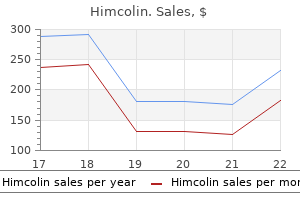
| Comparative prices of Himcolin | | # | Retailer | Average price | | 1 | Kroger | 133 | | 2 | Publix | 453 | | 3 | Delhaize America | 368 | | 4 | J.C. Penney | 289 | | 5 | Barnes & Noble | 980 | | 6 | Defense Commissary Agy. | 703 | | 7 | H-E-B | 822 | | 8 | Save Mart | 226 | | 9 | Kohl's | 463 |
Discount 30 gm himcolin with visaThis can lead to extreme cyanosis and hypoxia resulting in impotence due to alcohol generic himcolin 30 gm with amex metabolic acidosis erectile dysfunction prescription drugs himcolin 30 gm buy with mastercard, which is incompatible with life. In infants "double circles" in distinction to the traditional "circle and sausage" appearance. Newborns present with average to extreme cyanosis usu- ally in the first week of life. Cardiac catheter- ization and a balloon atrial septostomy (Rashkind procedure) are sometimes performed. The definitive correction is by the arterial switch operation, which is the process of alternative. Fontan Circulation the Fontan procedure is a palliative surgical procedure that redirects the systemic venous return on to the pulmonary arteries without passing through a subpulmonary ventricle. The complete cavopulmonary anastomosis or lateral tunnel Fontan consists of a direct, end-to-side superior cavopulmonary anas- tomosis (bidirectional Glenn operation) together with an intra- atrial baffle connecting the inferior vena cava to the underside of the pulmonary artery. The long-term issues embrace protein dropping enteropathy because of elevated venous pres- positive and intestinal lymphangiectasia, arrhythmias, throm- boembolic problems, obstruction of the conduit, and progressive ventricular dysfunction and cyanosis. What is a straightforward clinical check that can assist you to to differenti- ate between the 2 possible diagnoses Pulse oximetry between feeds his saturation drops to the 60s especially when he cries. Congenital Heart Defects: Decision Making for Cardiac Surgery: Volume 1 Common Defects. Two important determinants of blood pressure are cardiac output and whole peripheral resistance which shall be mentioned in detail later within the chapter. Hypertension is among the most typical circumstances, especially within the getting older popula- tion. It is often referred to as the silent killer as a outcome of it stays asymptomatic till it manifests as one of many life- threatening complications, for instance, stroke, myocardial infarction, kidney dysfunction, and so forth. The incidence of hypertension has been increasing in industrial international locations, which can be attributed to a large extent to dietary habits and an increase in weight problems. Furthermore, hypertension is an important worldwide public well being challenge- in a pooled data evaluate (Kearney et al. The variety of adults with hypertension is predicted to improve by 60% to a complete of 1. Factors that improve the guts price or stroke volume end in changes in blood strain. The stroke volume is dependent on cardiac contractility and blood quantity, which equates with sodium homeostasis. The autonomic nervous system affects both cardiac contractility and stroke volume. Another factor impacting cardiac output is the guts price that is also controlled primarily by the autonomic nervous system. Both environmental and genetic elements play an important role in blood stress control. Intracellular calcium has been associ- ated with increased muscle tone of the vascular smooth mus- cle and elevated blood strain. A calcium-rich diet has also been related to a reduction in the lipogenesis within the fats cells, thus providing an extra useful impact on blood strain. Other environmental components embrace low ranges of physical exercise, elevated stress, elevated levels of alcohol consumption, dyslipidemia, character traits (eg, a hostile perspective or time urgency, impatience) can negatively influence the blood pressure. An necessary genetic mutation concerned within the pathogenesis of hypertension is the Adducin family of genes (genes for cytoskeleton protein Adducin), which can result in elevated sodium reabsorption from the kidneys. The Connexin 40 gene mutation (a hole junction protein gene in the juxtaglomerular apparatus) has also been impli- cated in the pathogenesis of hypertension. Another necessary mutation is the angiotensinogen gene, which ends up in elevated levels of angiotensinogen, a precursor for angiotensin. Alto- gether, the interaction of both environmental and genetic factors determines the increased ranges of blood pressure. Stretch-sensitive sen- sory nerve root endings are situated in the carotid sinuses and the aortic arch. As the arterial stress rises, the rate of firing of these neurons increases, inflicting a decrease of sympathetic outflow, which in turn causes a decrease in the coronary heart rate and arterial stress. This is the first mechanism for the regu- lation of blood pressure in an acute setting, and acts because the buffer in changes of posture and acute changes within the blood volume. However, if the blood strain stays elevated, a downregulation of the baroreceptor reflex happens, and is ready to a better pressure level. Long-term blood strain main- tenance is dependent mainly on intravascular blood quantity via the renin-angiotensin-aldosterone mechanism. As the intravascular quantity will increase, the stroke quantity and cardiac output increases, and this causes the blood pressure to rise. However, if blood stress stays elevated for a protracted period of time, the whole peripheral resistance will decrease and the cardiac output will become normal. Primary versus Secondary Hypertension Primary hypertension, or hypertension without an identifiable cause (formerly called essential hypertension), accounts for approximately 95% of all cases of hypertension. Important factors that may contribute to this situation include elevated sympathetic activity and responsiveness of the adrenergic system. Increased angioten- sin 11 activity and mineralocorticoid excess are other impor- tant concerns. Primary hypertension is 4 occasions more frequent in African Americans and progresses extra rapidly, and is associ- ated with more complications as compared to charges of main hypertension in Caucasians. The pathophysiology behind age relates the increase in blood stress to the loss of elasticity, stiffening of the arteries, and a decrease within the renal capacity to hypertension and requires further workup (ie, diagnostic tests). Renovascular Hypertension Renovascular hypertension is the most typical explanation for sec- ondary hypertension and is potentially correctable. The former is common in older patients who generally produce other manifestations of atherosclerotic illness. Generally, athero- sclerotic plaque involves the proximal renal arteries at their origin in patients with atherosclerotic illness. Fibromuscular dysplasia is extra frequently associated with younger Cauca- sian females (8 occasions extra widespread than in different population groups). The particular selection of the check is decided by the condition of the affected person and the obtainable experience. Once a prognosis is established, treatment options depend upon patient traits and the goals of remedy. There is a powerful pathogenic association of insulin resis- tance with hypertension. When three of these 5 manifestations are present in a affected person, metabolic syndrome is diagnosed. Metabolic syn- drome increases the chance of heart disease, stroke, and diabetes mellitus. The precise mechanism by which insulin resistance induces hypertension remains to be unknown; however, insulin is known to improve each sympathetic exercise and sodium and water retention. This seems to be the most believable hypoth- esis for the correlation of insulin and hypertension.
30 gm himcolin cheap visaThe bladder is stuffed beneath gravity with 200 to 400 cc of distinction relying on bladder measurement and affected person consolation male erectile dysfunction pills review buy 30 gm himcolin. Adequate filling is essential to demonstrate intravesical pathology or bladder rupture erectile dysfunction doctors in maine generic 30 gm himcolin with amex. Oblique movies should be obtained because posterior diverticula or fistulae could additionally be obscured by the full bladder. The research supplies priceless data concerning the posterior urethra in pediatric sufferers. Evaluation of intravesical pathology of bladder diverticula of inguinal hernia involving the bladder of colovesical or vesicovaginal fistulae of bladder or anastomotic integrity after surgical of blunt or penetrating trauma to the bladder Technique the research could additionally be performed with the patient supine or in a semiupright place utilizing a desk able to bringing the patient into the total upright position. In children, a 5- to 8-Fr feeding tube is used to fill the bladder to the suitable quantity. Patient consolation should be taken into consideration when determining the appropriate quantity. A voiding cystourethrogram carried out for the analysis of recurrent urinary tract an infection in this female patient. Bladder filling in patients with spinal cord accidents greater than T6 may precipitate autonomic dysreflexia (Barbaric, 1976; Fleischman and Shah, 1977; Linsenmeyer et al. It is very delicate to adjustments that induce focal or world adjustments in kidney function. Compared with different diagnostic imaging studies corresponding to retrograde pyelogram, renal scintigraphy is noninvasive, has minimal risk and minimal discomfort, and allows dedication of the function of the kidney. Once the agent is intravenously injected, gamma scintillation cameras measure radiation emitted from the radioisotope and digital Indications 1. Evaluation of the urethra in men and women Limitations this study requires bladder filling utilizing a catheter. This could also be traumatic in kids and tough in some sufferers with anatomic abnormalities of the urethra or bladder neck. The tracer is properly suited for analysis of renal operate and diuretic scintigraphy. The preliminary section is the circulate section, by which 2-second pictures are gathered for 2 minutes and then 1-second pictures for 60 seconds. The circulate phase reveals renal uptake, background clearance, and abnormal vascular lesions, which may indicate arteriovenous malformations, tumors, or active bleeding. In the second part, the renal part, time to peak uptake is usually between 2 and four minutes. The renal section is the most delicate indicator of renal dysfunction; 1-minute pictures are taken for half-hour. In the ultimate phase, the excretory section, 1-minute pictures are taken for half-hour. The T1/2 is the time it takes for accumulating system activity to lower by 50% from that on the time of diuretic administration. This is very technician dependent because the diuretic must be given when the accumulating system is displaying maximum activity. Transit time through the amassing system in less than 10 minutes is in keeping with a traditional, nonobstructed amassing system. T1/2 of 10 to 20 minutes reveals mild to reasonable delay and could also be a mechanical obstruction. The stage of obstruction often could be decided as can abnormalities corresponding to ureteral duplication (Ell and Gambhir, 2004). The diuretic renal scan is one other imaging examine during which communication with the interpreting physician is important for correct efficiency of the test in addition to appropriate interpretation. For example, there are times when patients with unilateral or bilateral ureteral stents are despatched for diuretic scintigraphy to decide differential renal function. Dynamic perform images demonstrate good uptake of tracer by each kidneys and immediate visualization of the accumulating methods. Printout of quantitative data reveals the differential renal function to be 47% on the left, 53% on the proper. The T12 is 5 min on the left and seven min on the right, according to both kidneys being unobstructed. Gallbladder exercise, specifically, can cause false-positive interpretation when it overlies activity in the renal collecting system or is inappropriately included within the space of interrogation. Liver exercise is variable and tends to be more pronounced in kids and sufferers with renal insufficiency. This false-positive test might result in inappropriately reconstructing a kidney that has little or insufficient operate. Nuclear Medicine in Urologic Oncology Whole-Body Bone Scan Conventional radionuclide imaging in urologic malignancy has lengthy been the usual for detecting bone metastasis. The whole-body bone scan, or skeletal scintigraphy, is the most sensitive method for detecting bone metastasis (Narayan et al. This affected person introduced with a right-sided seminoma with cumbersome right-sided retroperitoneal lymph nodes. Tomography is an imaging methodology that produces 3D photographs of inner structures by recording the passage of x-rays as they move by way of completely different physique tissues. A collimated x-ray beam is generated on one facet of the patient and the quantity of transmitted radiation is measured by a detector placed on the other facet of the x-ray beam. These measurements are then repeated systematically while a series of exposures from totally different projections is made as the x-ray beam rotates around the affected person. Data collected by the detectors are reconstructed by computerized algorithms to lead to a viewable tomographic display. There are a quantity of totally different imaging variables which are adjusted to permit sufficient, detailed image decision, while minimizing the time on the scanner and limiting publicity to radiation. The variable software of pitch, beam collimation, detector size, and tube voltage are used by the radiologist and imaging technologist for best picture requisition. A detailed description of each of these variables is past the scope of this chapter. The helical raw images are processed using interpolation algorithms to visualize the inner structures as sagittal, coronal, or axial reconstructed images. These 3D photographs supply improved preoperative planning, appreciation of proximity to adjacent organs, and the flexibility to outline vasculature and enhance communication with sufferers who can now simply see their explicit pathology and better recognize the challenges confronted by their surgeon. It additionally has been used for fluid aspiration, drain placement, catheter placement, percutaneous cryoablation, and radiofrequency ablation of renal tumors. It is indicated within the workup of hematuria, kidney stones, renal masses, renal colic, and urothelial tumors. The thin slices offered by the 16-slice detector provide much greater detail of inner structures.
Himcolin 30 gm buy generic on linePreload Preload within the coronary heart can be outlined as the stretching of the myocardial muscle fibers simply prior to erectile dysfunction pumps side effects order himcolin 30 gm visa a contraction or ventricular wall pressure on the finish of diastole erectile dysfunction caused by herpes order himcolin 30 gm fast delivery. The importance of preload was described by Frank and Starling in an experimental mannequin, which confirmed that inside a physiologic vary, the more the ventricle is distended and crammed with blood during diastole, the extra the ventricle contracts, and more blood is and (3) a decreased Ca2+ inward present because of lowering useful Ca2+ channels. Thus, more depolarization is needed to reach the edge and fireplace an action potential. It is equivalent to the difference between the amount of blood within the ventricle just before the contraction (end-diastolic volume) and the vol- ume of blood left within the ventricle after the contraction (end- systolic volume). Stroke volume (mL) 2 End-diastolic quantity (mL) - End-systolic volume (mL) ejected within the next systole. Thus, inside physiological limits, the extra blood returning to the center as venous return, the extra blood pumped from the heart as cardiac output. It is affected by the neurotransmitters or hormonal influences and is principally mediated by the change of the intracellular calcium concentra- tion within the cardiomyocytes. In experimental studies, the primary index for contractility is often the change in pressure versus the change in time (dp/dt). However, in a medical setting, the index used for contractil- ity known as the ejection fraction. The ejection fraction is the fraction of the end-diastolic blood quantity ejected from 1 ven- tricle in 1 beat and calculated as follows: Ejection fraction 2 Stroke volume/End-diastolic volume 2 70/110 = 0. The impact of increased contractility on the stroke volume or the cardiac output will be mentioned in the Frank-Starling Relationship in following section. The activation of sympathetic exercise, for instance, dur- ing train or the administration of optimistic inotropic drugs, not solely leads to elevated cardiac contractility but additionally enhanced leisure. Overall, train or constructive inotro- Preload Relationship between preload and cardiac output. A clinical instance of a decreas- ing preload is a extreme hemorrhage or dehydration that can lead to a reduction of stroke quantity or cardiac output. Afterload Afterload is defined as the ventricular wall stress through the ejection part of systole or the resistance that the ventricle must overcome in order to eject its content material. Afterload is deter- mined by a number of components, including wall stress, aortic pres- sure, and whole peripheral resistance. Wall stress is the identical as the ventricular pressure (P), multi- plied by the ventricular radius (r), and divided by 2 times the wall thickness (h), as shown beneath. Wall pressure, on the opposite hand, is the same as wall stress but without consideration of pic medicine trigger the center to be more environment friendly throughout systole and diastole through (1) elevated dp/dt, thus growing the slope of the contraction and the speed of pressure development; (2) an increased peak within the left ventricular stress; (3) an increased rate of rest due to the enhanced rate of Ca2+ sequestration; and (4) a decreased systolic interval and more contractions during certain time intervals. Therefore, the cardiac output will increase to provide more blood provide to the lively muscles during exercise, or makes an attempt to return the cardiac output towards regular, for instance in coronary heart failure. Measurement of Cardiac Output There are a number of invasive and noninvasive strategies which are used to measure cardiac output. An invasive method includes intracardiac catheterization based on the conservation of mass utilizing the Fick principle. Briefly, by rearranging the next equations: 02 Consumption 2 Cardiac output X [02] Pulmonary vein - Cardiac output X [02] Pulmonary artery the wall thickness. Wall stress = (P X r) / 2h Wall stress is estimated from the law of Laplace as an increase within the ventricular pressure or the radius of the ventricle. However, elevated wall thickness reduces the wall stress as a outcome of the pressure is divided into a bigger thickness per unit of stress. For instance, a hypertrophied left ventricle acts as a compensatory mechanism for increased afterload and as a result reduces wall stress. Classic examples of increased afterload embody hypertension and aortic stenosis. Cardiac output 2 O2 Consumption/[02] Pulmonary vein - [02] Pulmonary artery 2 250 O2 mL/mirfl0. T-tubules form triads with the sarcoplas- mic reticulum to facilitate the release of Ca2+ throughout a contraction. Contractile Apparatuses within the Myocardium this section will concentrate on contractility of the myocardium, which is the muscular layer of the wall of the guts. Cardiac muscle is composed of numerous myocardial cells work- ing in conjunction with one another to pump blood through- out the physique. They consist of two types of membrane structures known as desmosomes and hole junctions. A desmosome is an adhering structure that mechanically aids to maintain adjoining cells together contemplating the excessive mechanical stress that happens in the myocardium. Gap junctions are low resistance paths that allow for the propagation of the electrical action poten- tial to adjacent cells. Initiation of an action potential leads to the activation of L-type Ca2+ channels on the myocyte membrane. When contraction happens, cross- bridges form between the actin and the myosin allowing adj a- cent filaments to slide throughout each other. This ends in a shortening of the sarcomere and thus the myofibril and the muscle contract. The 2 types of filaments involved in this pro- cess are thick and thin filaments. Thick filaments are com- posed of myosin and skinny filaments are composed of actin, troponin, and tropomyosin. Troponin is made up of three isoforrns, troponin T (TnT), troponin I (TnI), and troponin C (TnC). Rather, it binds to ryanodine channel recep- tors on the sarcoplasmic reticulum, which outcomes in the discharge � the depolarization of 1 myocardial cell leads to subsequent depolarization of the adjoining myocardial cells in an all or nothing style, meaning when 1 cell is electrically excited, an motion potential is propagated all through the entire myo- cardium. The term "electrical syncytium" is commonly used to por- tray this interconnectedness of how both the atria act as 1 unit and also how both the ventricles act as 1 unit. Structures such as the sarcoplasmic reticulum, T-tubules, and gap junctions facilitate this connection. The sarcoplasmic reticulum spans between the myofibrils and the T-tubular system, and the hole junctions are situated between the myocardial cells. This binding inhibits the action of troponin I and leads to a conformational change in the tropomyosin, freeing the myosin head binding site on actin. Energy obtained from the phosphate bond generates the "power stroke" that results in the movement of the thin filament over the thick filament inward and thus a myocyte contraction. Another necessary protein is phospholamban which is situated on the membrane of the sarcoplasmic reticulum. When phosphorylated, it results in the reuptake of cytosolic Ca2+ from the cytoplasm again into the sarcoplasmic reticulum, which results in leisure of the myocardial cell. In addition to the Ca2+ uptake into the sarcoplasmic reticulum, the NaJt-Ca2+ exchanger and the mebooksfree. Diagram of cardiac muscle cells indicates characteristic options of this muscle kind.
Order himcolin 30 gm onlineEven then erectile dysfunction virgin cheap 30 gm himcolin free shipping, the utility of percutaneous entry to the upper urinary tract collecting system was limited to drainage of obstructed kidneys till Fernstr�m and Johansson (1976) described the percutaneous removal of renal calculi erectile dysfunction pill brands himcolin 30 gm discount, termed percutaneous pyelolithotomy. Subsequently, many procedures have been performed by way of the percutaneous path to the upper urinary tract collecting system including drainage of an obstructed kidney, nephrolithotomy, endopyelotomy, and resection of urothelial tumors. More recently, percutaneous access to portions of the kidney apart from the collecting system has been introduced (addressed in Chapter 84). Specific elements of the procedures performed through the percutaneous access are lined in Chapters forty two, 86, and ninety nine. This is particularly true when the ureteral obstruction is long, extreme, or involving the ureteral orifice-which can retrograde access tougher. Conversely, untreated coagulopathy is a contraindication to percutaneous entry, however ureteral stents can be placed safely in an anticoagulated affected person. Studies utilizing a validated questionnaire in a inhabitants of patients after percutaneous nephrolithotomy counsel that a short lived nephrostomy tube, although equal to a ureteral stent when it comes to effectiveness of renal drainage, may actually be associated with improved health-related quality of life (Jiang et al. Diagnostic Studies In uncommon situations, percutaneous renal access is critical solely for diagnostic purposes, such as the Whittaker test, which is an invasive however highly accurate study to differentiate obstructive from nonobstructive hydronephrosis (Jaffe and Middleton, 1980). After obtaining percutaneous access into the kidney, the higher urinary tract is perfused at a continuing fee and pressures are transduced within the renal pelvis and urinary bladder. The discovering of a pressure differential above 12 cm of water across the ureteropelvic junction or ureter signifies urinary obstruction. Indications Simple Drainage Percutaneous renal drainage is efficient for most, if not all, obstructions together with those in intrarenal, ureteropelvic junction, or ureteral places. An alternative to percutaneous drainage is drainage via a ureteral catheter or stent placed in a retrograde trend (later in this chapter). This contains most cases of acute and continual ureteral obstruction with out infection (Rosevear et al. In the setting of upper urinary tract collecting system obstruction sophisticated by an infection, nonetheless, drainage is an emergency and in many such cases, percutaneous somewhat than retrograde drainage could also be finest (Ng et al. Percutaneous nephrostomy tubes and retrograde ureteral stents are generally equivalent in their capacity to resolve fever in patients with higher urinary tract obstruction and fever (Goldsmith et al. Retrograde ureteral stent placement typically requires regional or general anesthesia, whereas a percutaneous nephrostomy tube can be inserted under native anesthesia; this is a vital consideration in an unwell affected person. Because the percutaneous route has a larger preliminary success rate than the retrograde one in instances during which the amassing system is dilated, it may be most well-liked in a patient who needs rapid Therapeutic Instillations Percutaneous renal access could be carried out to facilitate instillations of chemotherapeutic brokers for upper-tract urothelial lesions or agents for chemolysis of renal stones. For a more comprehensive description of these matters, check with Chapters 43 and ninety nine. Percutaneous Renal Surgery Percutaneous renal access facilitates surgery for higher tract calculi, urothelial tumors, obstruction, and calyceal diverticulae and hydrocalyces. For a extra complete description of these surgical procedures, check with Chapters 42, forty three, and ninety nine. Anatomic Considerations With restricted visualization of the kidney and surrounding structures throughout normal percutaneous entry guided by fluoroscopy or ultrasonography, understanding renal and perirenal anatomy is important. Perirenal Anatomy the kidneys are well-protected, situated in the retroperitoneum, and surrounded by adipose tissue. The mobility of the kidneys is limited by short renal hilar vessels, although nephroptosis can happen, especially in skinny girls with a paucity of perirenal fats. This can be troublesome throughout percutaneous punctures with the affected person in a prone place. Additional visceral relations to the kidney include the adrenal glands (medial to the upper pole of both kidneys), the duodenum and gallbladder (anterior and medial to the proper kidney), and the tail of the pancreas (anterior and medial to the left kidney). Renal Parenchyma and Collecting System the renal parenchyma is composed of the cortex and the medulla. The cortex, outermost, contains the glomeruli and proximal and distal convoluted tubules. These are inverted cones (the base of which is superficial and the apex is deep) that comprise the loops of Henle and the collecting ducts, which coalesce on the apex of the pyramid into papillary ducts that open on the floor of the renal papillae. The columns of Bertin are invaginations of cortical tissue that encompass the renal pyramids except at their apices. The renal papillae drain into the minor calyces, that are probably the most peripheral parts of the intrarenal amassing system. The outermost wall of the calyx, into which the papilla is ready, is the calyceal fornix. There are 5 to 14 minor calyces in every kidney (mean of eight, with 70% of kidneys having 7 to 9 minor calyces) (Sampaio and Mandarim-de-Lacerda, 1988). Compound calyces are the rule within the higher calyceal group, are common within the decrease calyceal group, and are uncommon in the center calyceal group. Occasionally a minor calyx will open instantly into the renal pelvis without an intervening infundibulum. The compound calyces of the poles of the kidney are oriented dealing with their respective poles. Drainage of the higher pole into the renal pelvis is by a single midline infundibulum in most kidneys. Drainage from the decrease pole is by way of a single infundibulum in about one half of kidneys and in any other case by way of a series of paired anterior and posterior calyces. The center calyces typically are organized in a sequence of paired anterior and posterior calyces. In about two thirds of kidneys, there are two main calyceal systems-an upper and lower one-and the middle calyces drain into both or each systems. In the other one third of kidneys, the with the decrease poles lateral to the upper poles. The kidneys are additionally tilted 30 degrees off the frontal airplane, with the decrease poles anterior to the higher poles. Finally, the kidneys are rotated out of the frontal airplane as properly, with the lateral aspect of the kidney posterior to the medial facet, such that every kidney is rotated 30 degrees posteriorly from the renal hilum. This brings the upper pole calyces medial and superficial (dorsal) in relation to the lower pole calyces, which may have necessary implications for profitable renal access. The pleura can be violated throughout percutaneous entry into the higher pole of the kidney. The lung is above the eleventh rib, so direct lung harm is unlikely until the tenth intercostal area (superior to the eleventh rib) is used as the entry web site. The ribs curve inferiorly from medial to lateral, such that more parts of the kidney may be approached subcostally with a medial as opposed to a lateral entry web site. On the right side, the liver is anterior to the higher pole of the kidney and may prolong in some people to cover the whole anterior floor. Both the liver and spleen can extend lateral to the kidneys and are due to this fact at risk for damage with a lateral puncture into the kidney. This is especially true in conditions during which the liver or spleen are enlarged due to other medical conditions. The ascending and descending colon could be lateral and even posterior to the best and left kidneys, respectively. The distinction pertains to the center and lower calyceal system, which incorporates (in nearly all middle techniques and roughly one half of the decrease system) paired anterior and posterior minor calyces.
30 gm himcolin purchase mastercardIts renal clearance is proportional to creatinine clearance shakeology erectile dysfunction 30 gm himcolin cheap mastercard, and the half-life is 36 to forty hours in sufferers with normal renal perform erectile dysfunction tucson purchase 30 gm himcolin mastercard. Bz-blockades can precipitate vasoconstriction, which can result in the exacerbation of Raynaud phenomenon or a worsening of peripheral vascular illness. Abrupt withdrawal of B-blockers results in the precipitation of myocardial ischemia in patients with coronary artery illness. Bz-blockades can impair recovery from hypoglycemia in diabetic patients struggling an insulin response. This motion is respon- sible for the optimistic inotropic motion of digoxin, and likewise for the toxicity of the drug. Other molecular-level results of digitalis have been studied within the heart and are mentioned below. Receptors for cardiac glycosides that exist on the sodium pump have led to the assump- tion that an endogenous digitalis-like steroid, possibly ouabain or marinobufagenin, must exist. This group consists of cardiac glycosides, sympathomimetic brokers, and phosphodiesterase inhibitors. All these drugs improve calcium within the cardiac myocyte and improve actin and myosin interplay, thus bettering myocardial contractility. They cause a shift within the ventricular contraction curve (FrankStarling curve) in an upward direction (ie, it improves the stroke volume and cardiac output for a given filling pressure). Mechanical efiects � Digoxin increases contraction of the cardiac sarcomere by increasing the free calcium con- centration within the cardiomyocyte during systole. Digitalis is the genus name for the household of plants that pro- vide many of the medically useful cardiac glycoside, digoxin. Electrical efiects - the effects of digitalis on the electrical prop- erties of the guts are a mixture of direct and autonomic actions (Table 17. At greater concentrations, the resting membrane potential is lowered (made much less negative) because of inhibition of the sodium pump and decreased intracellular potassium. With further intoxication, each afterpotential-evoked action potential will itself elicit a suprathreshold afterpotential, and a self-sustaining tachycardia will be established. If allowed to progress, such a tachycardia can deteriorate into fibrillation; within the case of ventricular fibrillation, the arrhyth- mia might be rapidly deadly until corrected. This action involves sensitization of the baroreceptors, central vagal stimulation, elderly-and visible disturbances are noted. Interactions with Potassium, Calcium, and Magnesium Potassium and digitalis interact in 2 methods. Second, abnormal cardiac auto- and facilitation of the muscarinic transmission on the cardiac muscle cell. Cholinergic innerva- tion is much richer within the atria, so these actions have an effect on atrial and atrioventricular nodal operate more than the ventricles. Outside of the center, the gastrointestinal tract maticity is inhibited by hyperkalemia. Hypercalcemia increases the risk of a digitalis- induced arrhythmia by accelerating an overload of intracellular calcium shops that seems to be responsible for digitalis-induced abnormal automaticity. Therefore, serum electrolytes must be fastidiously moni- tored in sufferers on digoxin therapy. Less typically, disorien- tation and hallucinations-especially within the Clinical use Digoxin is reserved for treatment in patients with conges- tive coronary heart failure with atrial fibrillation as it has the good factor about ventricular fee management in this case. Milrinone increases myocardial contractility by increas- ing inward calcium flux within the heart. It can even alter the intra- mobile movement of calcium by influencing the sarcoplasmic reticulum. The impact zero Digitalis Toxicity zero the chance of digitalis toxicity is important because of the slender therapeutic index. The commonest cardiac manifestations of digitalis toxicity include atrioventricular junctional rhythm, premature ven- tricular depolarizations, bigeminal rhythm, ventricu- lar tachycardia, and second-degree atrioventricular blockade. The toxicity of inamrinone contains nausea and vomiting, arrhythmias, thrombocytopenia, and elevated liver enzymes. Milrinone is much less likely to trigger bone marrow and liver toxic- ity, but it could trigger arrhythmias. Milrinone is now used only intravenously and just for acute heart failure or extreme exac- erbation of persistent heart failure. In patients with extreme intoxica- tion, the administration of digoxin-specific antibodies could be life-saving. The improve in calcium entry trig- gers an increase in calcium release from the sarcoplasmic reticulum. Both intravenous dopamine and dobutamine are used incessantly within the remedy of acute heart failure. Norepi- nephrine, epinephrine, and isoproterenol are used in special circumstances. This combination of effects is due to its motion on completely different adrenergic receptors. It is utilized in remedy of coronary heart failure related to hypotension and impaired renal perfusion. At low doses (<2 ug/kg/min), dopamine activates primarily dopaminergic receptors in the renal and mesenteric vasculature; thus vasodilatation occurs with increased Other Positive lnotropic Drugs Used in Heart Failure Maj or efforts are underway to discover safer, constructive inotropic agents as a end result of cardiac glycosides have an especially slender therapeu- tic index and should not lower mortality in chronic heart failure. It has an elimination renal blood circulate and glomerular filtration with the facilitation of diuresis. Medium doses (2-10 ug/kg/min) have an inotropic action by stimulating cardiac Bl-receptors and indirectly promoting norepinephrine release from the sympathetic nerve terminals. This effect will increase the heart price, myocardial contractility, and stroke quantity, thus increasing the cardiac output. At excessive doses (>10 ug/kg/min), dopamine also stimulates oc-receptors, thus inducing vasoconstriction and rising systemic vascular resistance. Dobutamine is an artificial analog of dopamine that stim- ulates I31", [323 and oc-receptors. The drug is helpful within the treat- ment of heart failure not accompanied by hypotension. The drug is efficient for short length of treatment (<1 week), as a downregulation of the adrenergic receptors leads to much less results of dobutamine with extended use. The main antagonistic impact is tachyarrhythmias, though much less extreme than with dopamine. Norepinephrine is an endogenous catecholamine, syn- thesized from dopamine in adrenergic postganglionic nerves and in the adrenal medulla. Through its stimulatory impact on Bl-receptors, the drug has optimistic inotropic results. The enhance in peripheral vascular resistance will increase the mean arterial blood pressure. With this combined impact, norepinephrine is helpful in sufferers with "heat shock," during which heart failure is associ- ated with hypotension as a result of vasodilatation. It should be famous that vasoconstriction induced by norepinephrine is marked and might result in additional impairment in cardiac contractility, and are required.
|
