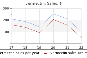
Cheap ivermectin 12 mg without prescriptionDecidua capsularis: Chorionic villi regress leaving a easy floor called chorion laeve antibiotics for uti and ear infection order 6 mg ivermectin. Decidua basalis: Chorionic villi are well developed (chorion frondosum) and contribute the fetal portion of the placenta antimicrobial resistance purchase ivermectin 12 mg amex. It seems on the dorsum (back) of the growing embryo, on the caudal (or posterior) end and proceeds towards the cephalic (anterior) end. It originates from the anterior epiblast, and seems as an elongating groove (primitive groove) on the dorsal midsagittal floor of the epiblast, alongside the anterior-posterior axis of the embryo. The rostrocaudal and medial-lateral axes of the embryo are outlined by the primitive streak. It can additionally be referred to as axial mesoderm) is an early forming midline structure in the trilaminar embryo mesoderm layer initially ventral to the ectoderm, then neural plate and at last neural tube. It is a transient embryonic anatomy structure, not current within the adult, defines the axis of embryo and is required for induction and Patterning the encompassing tissues. It types in week three, is eventually lost from vertebral regions and contributes to the nucleus pulposus of the intervertebral disc through the formation of the vertebral column. It varieties throughout gastrulation and soon after induces the formation of the neural plate (neurulation), synchronizing the event of the neural tube. Notochord formation: Epiblast cells on the floor of the amnion cavity in the blastopore region, kind a notochordal process, which later becomes notochordal canal and fuses with the endoderm to kind notochordal plate. This occurs on the ventral facet of the neural groove, the place an axial thickening of the endoderm takes place subsequent. This thickening seems as a furrow, the margins of which anastomose (come into contact), and so convert it right into a strong rod of polygonal-shaped cells (the definitive notochord) which is then separated from the endoderm. Notochord extends all through the whole length of the long run vertebral column, and reaches as far as the anterior finish of the midbrain, where it ends in a hook-like extremity within the area of the longer term dorsum sellae of the sphenoid bone. Initially it exists between the neural tube and the endoderm of the yolk-sac, however soon turns into separated from them by the mesoderm, which grows medially and surrounds it. From the mesoderm surrounding the neural tube and notochord, the skull, vertebral column, and the membranes of the brain and spinal cord are developed. Derived from hypoblast � Epiblast (not hypoblast) cells on the flooring of the amnion cavity in the blastopore region, kind a notochordal course of, which later evolve into definitive notochord. For instance, it induces the formation of the neural plate (neurulation), synchronizing the event of the neural tube. Nucleus pulposus � Notochord is a transient embryonic anatomy structure, which defines the axis of embryo. In the region of the node and streak, epiblast cells transfer inward (invaginate) to form new cell layers, endoderm and mesoderm. Hence, epiblast provides rise to all three germ layers in the embryo, ectoderm, mesoderm, and endoderm, and these layers form all of the tissues and organs. Prenotochordal cells invaginating within the primitive pit move forward until they reach the prechordal plate. With additional development, the plate detaches from the endoderm, and a solid wire, the notochord, is fashioned. Cephalic and caudal ends of the embryo are established before the primitive streak is fashioned. Normal L-R positioning of the organs is recognized as situs solitus, whereas their complete reversal is known as situs inversus. When a quantity of organs are abnormally positioned the situation is known as situs ambiguous or heterotaxy. Individuals with situs inversus have a low risk of getting other birth defects, however their kids have a higher threat, particularly for coronary heart defects. In distinction, sufferers with heterotaxy are at a excessive danger of having many types of congenital malformations, and virtually all will have some sort of cardiac abnormality. Epiblast cells moving by way of the node and streak are predetermined by their position to turn into specific types of mesoderm and endoderm. By the end of the third week, three primary germ layers, consisting of ectoderm, mesoderm, and endoderm, are established within the head region, and the method continues to produce these germ layers for more caudal areas of the embryo till the end of the fourth week. Tissue and organ differentiation has begun, and it occurs in a cephalocaudal course as gastrulation continues. When these villous capillaries make contact with capillaries within the chorionic plate and connecting stalk, the villous system is able to supply the embryo with its nutrients and oxygen. This course of is first indicated by the formation of the primitive streak within the midline of the epiblast. Caudal dysgenesis (sirenomelia or mermaid syndrome) is attributable to inadequate production of mesoderm by the primitive streak. Primitive streak � Primitive streak originates from the anterior epiblast, and appears as an elongating groove (primitive groove) on the dorsal midsagittal surface of the epiblast, along the anterior-posterior axis of the embryo. Primitive streak � Primitive streak is the groove formed in the epiblast on the caudal end of the bilaminar germ disc stage embryo by way of which epiblast cells migrate to form endoderm and mesoderm throughout gastrulation. Involves the hypoblast cells of inside cells mass � Gastrulation is the method by which the epiblast cells (not hypoblast) undergo ingression and establish three germ layers: endoderm, mesoderm and ectoderm. Hence the three germ layers are first seen close to the top region and consequently towards the tail region. Respiratory tract - epithelial lining, secretory and duct-lining cells of the trachea, bronchial, bronchloles and aveolar sacs. Epthelial lining, secretory and duct-lining cells of the oesophagus, abdomen and duodenum. Hepatocytes of liver, biliary tract, exocrine and endocrine cells of the pancreas. Midgut-epithelial lining, glandular and duct lining cells of the duodenum, jejunum, appendix, caecum, part of transverse colon Hindgut-epthelial lining, glandular and duct-lining cells of part of the transverse, decending and sigmoid colon, rectum, upper a part of anal canal. Allantosis-urinary bladder, vagina, urethra, secretory cells of the prostate and urethral glands. Coelomic wall epithelium Walls of intra-embryonic coelom Primitive pericardium - myocardium, parietal pericardium pericardioperitoneal canals - visceral, parietal and mediastinal pleura, pleuroperitoneal membranes contributing to diaphragm Splanchnopleuric epithelium - visceral peritoneum of abdomen, peritoneum of lesser and greater omental, faicliform ligament, lienorenal and gastrosplenica ligaments Somatopleuric epithelium - parietal peritoneum Primitive peritoneal cavity Splanchnopleuric epithelium - visceral peritoneal covering of mid- and hindgut, the mesentery, transverse and sigmoid mesocolon. Pronephros, epithelial lining of mesonephric ducts, vas deferens, epididymis, seminal vesicles, ejaculatory duct, ureters, vesical trigone Mullerian ducts, epithetical lining of uterine tubes, physique and cervix of uterus, vagina, broad ligament of uterus. Paraxial mesenchyme (somites and somitomeres) Sclerotome - vertebrae and parts of the neurocranium, axial skeleton Myotome - all voluntary muscles of the pinnacle, trunk and limbs. Intermediate mesenchyme - connective tissue of gonads, mesonephric and metanephric nephrons, samooth muscle and connective tissues of the reproductive tracts Septum transversum - epicardium, fibrous pericardium, portion of diaphragm, oesophageal mesentery, sinusoids of liver, tissue inside lesser omentum and falciform ligament. Lateral plate mesenchyme Splanchnopleuric layer - easy muscle and connective tissues of respiratory tract and associated glands. Smooth muscle and connective tissues of intestinal tract, associated glands and abdominal mesenteries. Smooth muscle and connective tissue of blood vessels (also see below) Somatopleuric layer - appendicular skeleton, connective tissue of limbs and trunk, including cartilage, ligaments and tendons.
Syndromes - Rapid heartbeat
- What other symptoms are present?
- Laxative
- Moving the person could cause further injury (for example, in case of a neck injury or motor vehicle accident)
- Lung volume can also be measured when you breathe nitrogen or helium gas through a tube for a certain period of time. The concentration of the gas in a chamber attached to the tube is measured to estimate the lung volume.
- Nephrotic syndrome
- Rigidity
- Bleeding into an area around the brain (also called a subarachnoid hemorrhage)
Ivermectin 12 mg order otcThe procedure consists of lifting the quadrate lobe upward (see [A] and [B]) antibiotic gram negative ivermectin 6 mg generic on-line, then not solely opening the umbilical fissure antibiotics for acne nausea discount ivermectin 3 mg otc, but also incising the deepest portion of the gallbladder fossa. At surgical pathology or 1-year follow-up, 55% have been noted to have viable tumor or recurrence. In the group determined to have a whole radiographic response, the subgroup of complete pathologic response has been reported between 17% and 65%. When sufferers with a whole pathologic response undergo resection, 5-year survival charges have been reported as much as 75%, by two unbiased groups. Five-year survival was 37%, 30%, and 8% for sufferers with objective tumor response, tumor stabilization, and tumor development, respectively. Control of metastatic illness earlier than surgical procedure may be crucial for an opportunity of extended remission in sufferers at excessive risk for progression after resection. If the anticipated functional hepatic volume after hepatic resection is taken into account marginal, strategies utilizing hepatic regeneration can rework some patients from unresectable to resectable. After hypertrophy of the remnant liver volume has plateaued, usually four to 6 weeks after the procedure, hepatic resection can be carried out. Some have advised that inducing liver hypertrophy might promote tumor development and lead to the next recurrence. Note the cystic plate (A) above the gallbladder, the hilar plate (B) above the biliary confluence and at the base of the quadrate lobe, and the umbilical plate (C) above the umbilical portion of the portal vein. Arrows point out the airplane of dissection of the cystic plate throughout cholecystectomy and of the hilar plate throughout approaches to the left hepatic duct. Philadelphia; Saunders; 2011; with laparoscopic video contributions from Carlos U. Colorectal liver metastases: disappearing lesions within the period of Eovist hepatobiliary magnetic resonance imaging. Indeed, response to chemotherapy before hepatic resection has turn into a serious choice factor for resection. Tumor stabilization or a lower in tumor burden throughout chemotherapy Two-stage hepatectomy consists of sequentially resecting hepatic metastases that may otherwise be unresectable because of insufficient hepatic reserve. This option is normally reserved for patients with multiple bilobar metastases aware of chemotherapy. The preliminary hepatic resection for metastases is carried out on the planned remnant liver, which allows it to hypertrophy within the absence of metastasis. The second hepatic resection for metastases is carried out after restaging at 6 weeks from authentic hepatectomy to enable the early regeneration of the remnant liver. Postoperative chemotherapy, consisting of the same proven chemotherapy that the affected person responded to beforehand, is continued for additional response. Of the 111 sufferers, some developed isolated hepatic recurrence (n = 40), isolated extrahepatic recurrence (n = 12), or both hepatic and extrahepatic recurrence (n = 59). Isolated hepatic recurrences have been handled by repeat hepatectomy, whereas patients with extrahepatic tumors underwent resection. Overall remedy of the 138 patients included 223 hepatectomies, forty two particular procedures to enable resectability, and seventy seven extrahepatic surgical procedures. Patients whose hepatic metastases had been initially resectable had 3-, 5-, and 10-year overall survival of 66%, 48%, and 30%, respectively. Downstaging of metastatic illness and modalities to induce selective hypertrophy of the remnant liver can now permit hepatic resection with healing intent in selective responsive sufferers. Coagulation profiles (rarely needed) are corrected, and prophylactic antibiotics directed at higher gastrointestinal tract flora are administered at the time of surgery. Intraoperative management of the patient is important to the success of the process. The maintenance of low central venous strain (5 to 7 mm Hg) reduces parenchymal blood loss by way of hepatic venous sources. The major pitfalls or hazard points with hepatic resection embrace hemorrhage from hepatic veins, portal veins, or hepatic arteries; air embolism from hepatic venous harm; damage to the biliary ductal system with postoperative obstruction or fistula formation; portal or hepatic vein compromise with subsequent ischemia or postsinusoidal portal hypertension; prolonged vascular influx occlusion resulting in refractory hepatic ischemia or hepatic injury; and injury to the diaphragm, inferior vena cava, or gut. The liver is mobilized by full division of the suitable ligamentous attachments. The skinny gastrohepatic omentum is incised adjacent to the hepatoduodenal ligament. The foramen of Winslow is opened in anticipation of subsequent influx vascular occlusion. In our follow, a Thompson retractor is used to elevate the rib cage anteriorly and cephalad while retracting the remaining viscera caudally. Bile ducts or vessels with diameter bigger than 2 mm are clamped with metallic clips or ligated with suture. Suture ligation of remnant vessels or ducts reduces artifacts during postoperative imaging. Closed, low-pressure suction drainage is optionally available and tends to be used based on surgeon choice. The current suggestions for formal terminology have been proposed and are referenced for review. Wedge resections are normally carried out with a minimum of blood loss, usually without inflow vascular occlusion. Portal and segmental pedicles are greatest approached by dissection from the hilus to the suitable pedicle or by direct rapid parenchymal transection along an estimated intersegmental plane with ultrasound steering. Both approaches are facilitated by momentary inflow vascular occlusion to reduce hemorrhage and by method of the ultrasonic aspirator to quickly expose the pedicles via the intervening parenchyma. Alternatively, some have proposed injection of methylene blue into the segmental or portal pedicle utilizing ultrasound steering, which provides visible identification of the anatomic segmental or sectoral anatomy. If so, the infrahepatic suprarenal and suprahepatic inferior vena cava are encircled in the course of the preliminary dissection to permit occlusion by vascular clamps or tapes. The portal triad to the proper liver has been divided, and a line of demarcation has developed. The parenchymal transection is done with a vessel loop occluding the hilar vessels (Pringle maneuver). The danger of blood loss is decreased considerably by ligation of the appropriate lobar hepatic arterial and portal venous branches before parenchymal transection. In addition, ligation of the corresponding hepatic vein before parenchymal transection to expose vessels or ducts for ligation or cauterization. Hemorrhage is decreased by digital compression of the liver on both sides of the transection plane. The hilar plate is lowered to expose the left hepatic duct and the confluence of the bile ducts. Hilar lymph nodes are excised to further expose the bile duct, portal vein, and hepatic artery and for staging.
Safe 6 mg ivermectinManagement of surgical and radiation induced rectourethral fistulae with an interposition muscle flap and selective buccal mucosal onlay graft infection 12 mg ivermectin buy with mastercard. Rectal injury occurring at radical retropubic prostatectomy for prostate most cancers: etiology and therapy antibiotics for acne bactrim ivermectin 3 mg generic mastercard. Urinary fistulae following exterior radiation or permanent brachytherapy for the remedy of prostate cancer. Rectal damage throughout robotassisted radical prostatectomy: incidence and administration. The incidence and administration of rectal injury related to radical prostatectomy in a community based urology follow. Incidence, clinical signs and management of rectourethral fistulae after radical prostatectomy. Rectourethral fistulae secondary to prostate most cancers therapy: management and outcomes from a multi-institutional combined experience. Actuarial disease-free survival after prostate most cancers brachytherapy utilizing interactive strategies with biplane ultrasound and fluoroscopic steerage. The feasibility and security of high-intensity focused ultrasound as salvage therapy for recurrent prostate cancer following external beam radiotherapy. Patient selection, most cancers management, and complications after salvage native remedy for postradiation prostate-specific antigen failure: a systematic review of the literature. Salvage prostate cryoablation: preliminary outcomes from the cryo on-line data registry. Long-term artificial urinary sphincter outcomes following a prior rectourethral fistula repair. High-dose rate interstitial with exterior beam irradiation for localized prostate cancer-results of a prospective trial. Surgical administration of complicated rectourethral fistulae in irradiated and nonirradiated patients. Successful restore of iatrogenic rectourinary fistulae utilizing the posterior sagittal transrectal approach (York-Mason): 15-year expertise. Suggested contributing factors embody standing, constipation, colic, coughing, sneezing, weak anal sphincters and pelvic floor musculature, an irregular cul-de-sac, and distal rectal intussusception. The main presenting criticism is commonly of a protuberance or bulge from the anus, which is usually mistaken for hemorrhoidal illness. Other complaints may be a feeling of incomplete evacuation of stool, bleeding, mucus discharge, urinary incontinence, and pain. The prolapse may be apparent on initial exam; different instances it will need to be elicited by asking the patient to carry out a Valsalva. Colonoscopy is required in all patients considered candidates for operative restore of rectal prolapse to rule out a neoplastic lead point. Other diagnostic tests of debatable significance embody anal manometry, endorectal ultrasound, and defecography. If important full-thickness ischemia is current, discount should be prevented and operative resection will probably be required. The majority of patients with viable prolapse will reply to continuous, regular strain within the workplace setting or emergency room. At instances, intravenous sedation or even basic anesthesia within the working room could additionally be needed for full discount. Applying desk salt or sugar to the mucosa can scale back the swelling of the incarcerated rectum and help in reduction. The transabdominal approach is the preferred repair in patients who can tolerate a transabdominal operation. Care must even be given to evaluating the affected person for the presence of concurrent anterior compartment prolapse to assess the feasibility of repair of those prolapsing organs as well in a combined operation. There are two primary technical parts within the belly method to rectal prolapse: rectopexy and resection. In 1963, Ripstein described the posterior rectopexy: a posterior rectal dissection to the level of the coccyx and a sling of fascia lata around the rectum anchored to the proximal sacrum. This method, nonetheless, results in barely larger recurrence rates than rectopexy with mesh (Table 152. The extent of rectal mobilization required throughout rectopexy has also been an issue of debate. In addition, it describes the nonoperative and operative administration choices of each situations intimately. Rectal prolapse has myriad stomach and perineal operative approaches related to increasingly comparable results. Conversely, inside rectal intussusception, until an isolated finding in a patient with defecatory dysfunction, stays largely a condition of nonoperative management. The most recent addition to the belly operations for rectal prolapse is the ventral rectopexy that includes mobilization of the anterior portion of the rectum and not utilizing a posterior or lateral dissection. Mesh formed in the type of a "hockey stick" is then sutured from the anterior rectal wall to the sacral promontory and coated with peritoneum. Some authors report lower constipation rates, while sustaining recurrence charges much like posterior rectopexy (see Table 152. The integrity of the repair has not been compromised with the laparoscopic strategy, as recurrence charges are related. The short-term postoperative benefits of laparoscopic surgical procedure are realized in patients with rectal prolapse, including decreased postoperative pain, shorter hospital size of stay, and faster return of bowel perform. Many of those sufferers have vital comorbidities that restrict their capability to tolerate an belly approach. In these circumstances, a perineal has been proven to improve the rate and severity of constipation. Modified perineal stapled rectal resection with Contour Transtar for full-thickness rectal prolapse. However, a perineal resection-rectopexy can spell doom if the blood provide of the rectosigmoid was previously divided, and the perineal resection leaves an ischemic segment of rectum in place. The anal canal is held open with a Lone Star retractor (Lone Star Company, Frisco, Texas). The avascular intersphincteric aircraft is developed as much as the pelvis, and the distal rectum is pulled from the pelvis. Using both suture ligation or an vitality system, the vascular supply (first laterally, then posteriorly) of the uncovered bowel is then ligated because the dissection is continued proximally. The redundant rectum and sigmoid colon are extracted to the extent of the left colon, and the bowel is divided underneath delicate stretch at the point of deliberate coloanal anastomosis. A posterior rectopexy with a postanal levatorplasty has been shown to improve anal continence. However, some authors report a better likelihood of recurrence with a stapled anastomosis. Using electrocautery, a circumferential submucosal incision is then made 1 cm proximal to the dentate line. Being careful not to violate the muscular layer of the rectal wall, a submucosal dissection is carried out and extended proximally. After the mucosal sleeve is excised, longitudinal plicating sutures are positioned in rows alongside the length of the rectal wall musculature 1 cm apart.
Buy 3 mg ivermectin visaLiver resection for metastatic colorectal most cancers in the age of neoadjuvant chemotherapy and bevacizumab treatment for dogs eating onions purchase 12 mg ivermectin fast delivery. Clinicopathological features and prognosis in resectable synchronous and metachronous colorectal liver metastasis antibiotics for uti cipro dosage ivermectin 6 mg without a prescription. Haematogenous metastatic patterns in colonic carcinoma: an analysis of 1541 necropsies. Visualizing portal vein metastatic trafficking to the liver with green fluorescent protein-expressing tumor cells. Managing synchronous liver metastases from colorectal most cancers: a multidisciplinary worldwide consensus. Simplified staging system for predicting the prognosis of patients with resectable liver metastasis: development and validation. Perioperative management of hepatic resection toward zero mortality and morbidity: analysis of 793 consecutive cases in a single establishment. Comparison of simultaneous or delayed liver surgical procedure for restricted synchronous colorectal metastases. Extension of the frontiers of surgical indications within the treatment of liver metastases from colorectal most cancers: long-term outcomes. Pathologic response to preoperative chemotherapy in colorectal liver metastases: fibrosis, not necrosis, predicts consequence. Tumor development whereas on chemotherapy: a contraindication to liver resection for multiple colorectal metastases Optimal morphologic response to preoperative chemotherapy: an alternate outcome finish level earlier than resection of hepatic colorectal metastases. Margin standing stays an important determinant of survival after surgical resection of colorectal liver metastases in the period of contemporary chemotherapy. Colorectal cancer liver metastases and concurrent extrahepatic disease treated with resection. Tumour biology of colorectal liver metastasis is a more necessary consider survival than surgical margin clearance within the period of contemporary chemotherapy regimens. Bevacizumab improves pathologic response and protects against hepatic harm in patients handled with oxaliplatin-based chemotherapy for colorectal liver metastases. Optimizing resection for "responding" hepatic metastases after neoadjuvant chemotherapy. Selection of patients for resection of hepatic colorectal metastases: expert consensus statement. Optimal future liver remnant in sufferers treated with in depth preoperative chemotherapy for colorectal liver metastases. Kinetic growth price after portal vein embolization predicts posthepatectomy outcomes: towards zero liver-related mortality in sufferers with colorectal liver metastases and small future liver remnant. Liver useful reserve estimation: state of the art and relevance for local treatments: the Eastern perspective. Assessment of future remnant liver function utilizing hepatobiliary scintigraphy in sufferers present process major liver resection. Use of scientific rating to stage and predict outcome of hepatic resection of metastatic colorectal cancer. Comparison of clinical threat scores predicting prognosis after resection of colorectal liver metastases. Influence of resection margin on survival in hepatic resections for colorectal liver metastases. Genetic and histological assessment of surgical margins in resected liver metastases from colorectal carcinoma: minimal surgical margins for profitable resection. Improving resectability of hepatic colorectal metastases: professional consensus assertion by Abdalla et al. Complete response of colorectal liver metastases after chemotherapy: does it mean treatment Microwave ablation: comparison of simultaneous and sequential activation of a number of antennas in liver mannequin systems. Evolution of surgical microwave ablation for the therapy of colorectal cancer liver metastasis: evaluation of the literature and a single centre expertise. Microwave ablation for unresectable hepatic tumours: medical outcomes using a novel microwave probe and generator. Procedures of choice for resection of major and recurrent liver metastases from colorectal most cancers. Resection of nonresectable liver metastases from colorectal most cancers after percutaneous portal vein embolization. Oncological outcomes of main liver resection following portal vein embolization: a scientific review and meta-analysis. Rescue surgical procedure for unresectable colorectal liver metastases downstaged by chemotherapy: a mannequin to predict long-term survival. Central venous stress and liver resection: a scientific review and meta-analysis. Regional chemotherapy: a concentrate on hepatic artery infusion for colorectal most cancers liver metastases. Intraarterial infusion chemotherapy for hepatic carcinoma using a totally implantable infusion pump. Technical problems and sturdiness of hepatic artery infusion pumps for unresectable colorectal liver metastases: an institutional experience of 544 consecutive instances. Hepatic arterial infusion of chemotherapy after resection of hepatic metastases from colorectal cancer. Reappraisal of hepatic arterial infusion in the treatment of nonresectable liver metastases from colorectal cancer. Conversion to full resection and/or ablation utilizing hepatic artery infusional chemotherapy in patients with unresectable liver metastases from colorectal cancer: a decade of expertise at a single establishment. Resection of the liver for colorectal carcinoma metastases: a multi-institutional examine of patterns of recurrence. Radiofrequency ablation for colorectal liver metastases: prognostic elements in non-surgical candidates. Five-year survival in 309 sufferers with colorectal liver metastases treated with radiofrequency ablation. Small liver colorectal metastases treated with percutaneous radiofrequency ablation: local response rate and long-term survival with 66. Malignancies of the anal canal and perianus are unusual and account for 2% of all lower gastrointestinal tract cancers. On digital examination, this corresponds to the palpable anorectal ring superiorly and the intersphincteric groove, the outermost boundary of the internal sphincter inferiorly. This is considered to be a variant of squamous epithelium and consists of cloacogenic, transitional, and basaloid epithelium as an alternative of the columnar epithelium of the rectum.
Ivermectin 3 mg buy fast deliveryThe terminal portion of mesodenphric duct (mesoderm) are absorbed into the posterior wall of urogenital sinus to type trigone the terminal portion of mesonephric duct will get absorbed into the posterior wall of urinary bladder to type the trigone (mesodermal) antibiotic joint replacement dental 6 mg ivermectin discount free shipping. Note: Proximal a half of mesonephric duct (Wolffian duct) kind the conduit for sperm from the testes to the urethra and provides rise to epididymis bacteria diagram 6 mg ivermectin sale, ductus deferens, seminal vesicle, common ejaculatory duct. Table 22: Development of male and female urethra Urethra Female urethra Male urethra 1. Male urethra Clinical Correlations Renal agenesis occurs when the ureteric bud fails to develop, thereby eliminating the induction of metanephric vesicles and nephron formation. Pelvic kidney is an ectopic kidney that occurs when kidneys fail to ascend and thus remain in the pelvis. Horseshoe kidney develops on account of fusion of the decrease poles of two kidneys. Urachal fistula or cyst happens when a remnant of the allantois persists, thereby forming fistula or cyst. It is found alongside the midline on a path from the umbilicus to the apex of the urinary bladder. A urachal fistula forms a direct connection between the urinary bladder and the surface of the physique at the umbilicus, causing urine drainage from the umbilicus. The transitional epithelium lining the urethra and the bladder is derived from: a. Connecting tubule is a derivative of ureteric bud � Connecting tubule is a part of nephron and develops from metanephric blastema. Note: Ureter develops from ureteric bud, which can be referred to as as metanephric diverticulum by some authors. Mesonephric duct � Mesonephric duct provides the ureteric bud which later develops into ureter. Supernumerary arteries � the most typical aberration in renal vessel improvement is supernumerary (accessory) renal arteries. Common ejaculatory duct � In each sexes, the mesonephric duct gives origin to the ureteric bud, which varieties the ureter, renal pelvis, main and minor calyces and the amassing tubules of the kidney. Endoderm � Urinary bladder develops from the endoderm of vesicourethral canal (cranial a part of urogenital sinus). Urachal fistula � Non-obliteration of allantois (hindgut diverticulum) might lead to patent allantoic (urachal) fistula, which results in leakage of urine from the urinary bladder in the course of the umbilicus, especially on straining. Inferior mesenteric artery � the conventional ascent of the kidneys allows the organs to take their place in the stomach under the adrenal glands. Mesonephros � Epithelium of ureter develops from the mesonephric duct, because the ureteric bud. Rectum and unogenital sinus � Urorectal septum divides the cloaca region into urogenital sinus (anterior) and rectum (posterior) 17. Mesonephric duct � the terminal portion of mesonephric duct gets absorbed into the posterior wall of urogenital sinus and types the trigone of urinary bladder. Patent allantois � Allantois gets obliterated to kind urachus, which may remain patent to type urachal fistula. Primordial germ cells originate in the epiblast cells (primitive streak), migrate to the endodermal wall of yolk sac and thence to the genital ridge (during the 4th to 6th weeks). During the indifferent stage, there are two duct systems: the mesonephric duct and paramesonephric duct. Testosterone, produced by Leydig cells in the testes, stimulates improvement of the mesonephric ducts to kind the efferent ducts, epididymis, vas deferens, seminal vesicle and ejaculatory duct. M�llerian inhibiting substance (Anti Mullerian hormone), produced by Sertoli cells in the testes, causes regression of the paramesonephric (Mullerian) ducts. Dihydrotestosterone stimulates growth of the exterior genitalia, together with the penis and scrotum. Estrogens (together with the absence of testosterone) regulate growth of the paramesonephric ducts, which finally ends up in genesis of the uterus, uterine tube and higher 1/3 of the vagina. Estrogens also stimulate differentiation of the external genitalia, together with the clitoris, labia, and lower portion of the vagina. Initially, a genital tubercle, two genital swellings, and two cloacal folds type on the outside of the ground of the pelvis. When the urorectal septum reaches the interior of this ground to separate the anal canal from the primitive urogenital sinus (soon to type the bladder), the cloacal folds at the second are referred to as the urethral folds. In females, the genital tubercle types the clitoris, the urethral folds the labia minora, and the genital swellings the labia majora. Mesenchyme Leydig cells (interstitial cells) Theca cells (forming theca interna and externa) 2. Round ligament of uterus 121 Self Assessment and Review of Anatomy Adult derivatives Embryonic construction three. Mullerian tubercle Genital tubercle Urethral folds Genital swellings Uterine tubes Uterus Cervix Upper a half of vagina Urinary bladder Urinary bladder Urethra (prostatic membranous and penile) Urethra (membranous) and vestibule of vagina Prostate gland Paraurethral glands (of Skene) Bulbourethral glands Greater vestibular glands Seminal colliculus (verumontanum) Hymen Penis Clitoris Penile urethra Labia minora Scrotum Labia majora Flowchart 5: Genetic basis of phenotypical differentiation of testis and ovary Flowchart 6: cells on indifferent gonad Table 24: Differences in improvement of testis and ovary Testis Formation of just one generation of intercourse cords (medullary cords) that produce seminiferous tubules and rete testis Ovary Formation of two generations of sex cords: a. Second era of intercourse cords (cortical cords) kind primordial follicles (ovarian follicles) No formation of tunica albuginea. Gubernaculum is the embryonic buildings which begin as undifferentiated mesenchyme attaching to the caudal end of the gonads (testes and ovaries). The testes descend to a larger diploma than the ovaries and finally move via the inguinal canal. In males the upper part of the gubernaculum degenerates and decrease part persists as the gubernaculum testis (scrotal ligament), which secures the testis to the most inferior portion of the scrotum, tethering it in place and limiting the degree to which the testis can transfer throughout the scrotum. High Yield Points Primordial germ cells are derived from the epiblast cells, which later migrate to the endodermal wall of yolk sac and eventually attain the genital ridge. Between the third and fifth gestational weeks, an elevation of intermediate mesoderm on both sides of the fetus - the urogenital ridge begins growth into the urogenital tract. There are 7 million oocytes at 5th month of intrauterine life, 2 million at birth and about four hundred bear ovulation. Upper a half of genital tube develops from intermediate mesoderm, whereas lower part from endoderm of urogenital sinus. Upper 1/3 vagina develops from Mullerian duct (intermediate mesoderm), whereas lower vagina develops from endoderm of urogenital sinus. The Wolffian duct give rise to epididymis, ductus deferens, seminal vesicle and customary ejaculatory ducts. Mesonephric or Wolffian vestiges can persist as Gartner duct cysts (anterolateral wall of vagina). Organ of Rosenm�ller (Epoophoron) is a remnant of the mesonephric tubules current near the ovary and fallopian tube. The appendix testis (hydatid of Morgagni) is a vestigial remnant of the Mullerian duct, attached to the upper pole of the testis. It is inconceivable to visually differentiate between female and male exterior genitalia until week 12. From 28 weeks the testes pass by way of the superficial inguinal ring and thence to scrotum. The major sex cords extend into the medulla of the gonad and lose their connection with the surface epithelium as the thick tunica albuginea types. Mesoderm between the seminiferous cords gives rise to the interstitial (Leydig) cells, which secrete testosterone.
Brickellia. Ivermectin. - Are there safety concerns?
- Dosing considerations for Brickellia.
- What is Brickellia?
- How does Brickellia work?
- Are there any interactions with medications?
- Diabetes, diarrhea, stomach pain, gallbladder disease, and other conditions.
Source: http://www.rxlist.com/script/main/art.asp?articlekey=97089
Ivermectin 12 mg discount mastercardLateral ligament division throughout rectopexy causes constipation however prevents recurrence: results of a prospective randomized study antibiotics for acne safe for pregnancy 3 mg ivermectin buy with visa. Surgery for rectal prolapse: Orr-Loygue ventral rectopexy with restricted dissection prevents postoperative-induced constipation without rising recurrence 7dtd infection purchase 6 mg ivermectin with amex. A prospective randomized research of belly rectopexy with and with out sigmoidectomy in rectal prolapse. Laparoscopic ventral recto(colpo)pexy for rectal prolapse: surgical technique and end result for 109 patients. Laparoscopic ventral rectopexy for exterior rectal prolapse improves constipation and avoids de novo constipation. Laparoscopic ventral rectopexy: a prospective long-term analysis of functional outcomes and quality of life. Laparoscopic-assisted resectionrectopexy for rectal prolapse: early and medium follow-up. Open vs laparoscopic repair of full-thickness rectal prolapse: a re-meta-analysis. Perineal proctectomy, posterior rectopexy, and postanal levator restore for the remedy of rectal prolapse. Long-term efficacy of biofeedback remedy for dyssynergic defecation: randomized managed trial. In patients with rectoceles and obstructed defecation syndrome, surgical procedure ought to be the option of final resort. Stapled transanal rectal resection for obstructed defecation and evidence-based follow. Functional results of laparoscopic resection rectopexy for symptomatic rectal intussusception. Obstructive defecation syndrome: 19 years of expertise with laparoscopic resection rectopexy. The present understanding is that that is an acquired illness leading to subcutaneous trauma of hair shafts. This idea motivated the Karydakis flap, wherein the sinus is excised and the intergluteal skin is changed by extra "resistant" gluteal skin via a sliding flap. Acute abscess is the presenting discovering in approximately 50% of sufferers and constitutes 20% of dermatologic complaints seen in the emergency division. In 1935, Gage proposed that sinus disease was secondary to anomalous embryonic improvement of the medullary canal. Wound issues and recurrence remain excessive despite obtainable therapies, making this a difficult illness course of for both the patient and surgeon. Current understanding is that this is an acquired course of secondary to occlusion of the apocrine ducts with secondary an infection. Medical remedy is beneficial for mild illness and is a helpful adjunct for symptom management. Acute Pilonidal Abscess Acutely, simple incision and drainage is the standard remedy to provide immediate symptom aid. In patients with acute abscess formation, a staged method must be employed as definitive resection with abscess excision leads to larger charges of recurrence. Drainage procedures may be carried out within the workplace, emergency room, or an outpatient ambulatory surgical setting. The sacrococcygeal region is finest exposed in the prone jackknife position with the buttocks taped aside. Unroofing and fulguration of all sinus tracts is supported even within the acute section with a healing time of 5. Weekly wound checks are beneficial to ensure that the wound and surrounding pores and skin remain free of hair and other particles during healing. Laser hair removal may be supplied as an adjunctive therapy during wound therapeutic, in addition to a long-term approach to reducing hair progress in the concerned area. Armstrong and Barcia27 advocated nonexcisional remedy consisting of meticulous hair control by shaving, good perineal hygiene, and limited lateral incision and drainage of abscesses. The microbiology of the pilonidal sinus is often gramnegative and anaerobic organisms, with a shift toward gram-positive and aerobic bacteria in recurrent disease. When the congenital principle prevailed, procedures were guided by the premise of complete extirpation of subcutaneous tissue to the extent of the sacral fascia. Pilonidal disease: origin from follicles of hairs and results of follicle elimination as treatment. Instillation of liquid or crystalline phenol causes an intense inflammatory reaction to promote closure of the cavity and tracts. This method has restricted use due to ache and should require hospitalization for analgesia. Midline follicle excision and lateral drainage was described by Bascom7 and an analogous technique described by Lord and Millar39-41 within the mid-1960s. This process includes a longitudinal incision off of midline for entry to the continual sinus tract. The sinus tract is d�brided of necrotic materials, hypergranulation tissue, and hairs. The space is opened within the midline, adopted by curettage of any particles or granulation tissue. Alternatively the skin edges can be sutured (marsupialization) to the base of the wound. Excision of the pilonidal cavity and associated inflammatory tissue is an extirpative method. Meta-analysis of 18 randomized managed trials compared the results of open versus closed surgical therapy for pilonidal sinus. The knowledge suggest extra speedy healing after primary closure when compared with excision with out closure. In addition, there was no difference in infection rates when evaluating the two strategies. There was a trend toward decreased recurrence after open therapeutic as in contrast with major closure; nevertheless sufferers did return to work faster after major closure. When comparing midline closure versus off-midline closure, there was good proof of slower healing, larger rates of infection, higher rates of recurrence, and different problems after midline major closure in contrast with off-midline closure strategies. In addition, recurrence charges have been reported to be wherever between 2% and 11%. Bascom described this technique for a continual nonhealing midline wound whereby the midline nonhealing wound is excised and a full-thickness skin flap is raised to overlap the skin edges of the wound on the other side. From a technical standpoint, this is considered one of the simpler flap procedures and can be carried out in an outpatient ambulatory surgical setting.
Generic ivermectin 12 mg free shippingPatients with troublesome colons due to antibiotic resistance lesson plan ivermectin 12 mg cheap mastercard acute angulation might benefit from the use of a pediatric colonoscope or even a gastroscope-both of these have a smaller diameter and tighter turning radius than an grownup colonoscope antibiotic yeast infection symptoms 3 mg ivermectin cheap with visa. The portion of the scope that connects from the deal with to the processor should also be positioned without any looping. Inspection of the perianal pores and skin is performed, followed by an intensive digital rectal examination. The left colon is the most tough a part of the colon to traverse within the majority of patients. Ideally, direct visualization of the lumen is maintained all through the process, using a mixture of torque on the insertion tube and angulation of the tip. Although clockwise torque is the predominant path utilized, there are some circumstances during which counterclockwise torque shall be wanted. Looping of the scope is frequent in the left side of the colon, however efforts to cut back looping ought to be made any time a relatively straight portion of the colon is encountered. A mixture of gradual withdrawal, aspiration of air, and torque all carried out concurrently while keeping the lumen in view with the angulation knobs is an effective loop-reducing method. Abdominal pressure, use of an adjustable stiffness colonoscope, and positional modifications are different adjuncts to allow for profitable cecal intubation. It should be careworn that cecal intubation means coming into into the cecum, proximal to the ileocecal valve, and not simply visualizing the appendiceal orifice when the scope is distal to the valve. Thorough inspection of the cecum requires distending it and pulling back on the scope while torqueing or angulating the tip downward to expose the realm immediately proximal to the valve-in this area aspiration of fuel may be necessary. When a affected person on antithrombotic therapy requires colonoscopy, the endoscopist should contemplate several components, such because the urgency of the process, bleeding risk related to the procedure, effects of the antithrombotic medicine on the bleeding dangers, and danger of stopping the antithrombotic therapy (Abraham bleeding threat and strategies). There are knowledge to help the utilization of chilly snare somewhat than sizzling snare in those sufferers anticoagulated with warfarin. In basic, the morbidity of a thromboembolic occasion is larger than the danger of hemorrhage. Therefore resuming anticoagulation as soon as attainable following colonoscopy is our present practice. Mucosal vessels highlighted due to absorption of green and blue wavelengths in hemoglobin Computerized digital manipulation of photographs utilizing wavelength combinations Computerized enhanced photographs of the mucosal surface and the blood vessels through postimage processing this will develop the ability so that the endoscopist is in a position to perform it when wanted. Retroflexion within the cecum can be performed and is sometimes necessary to take away ascending colon polyps. Thorough examination of all mucosal surfaces requires cleaning the colon wall with water and aspirating swimming pools of liquid. Skillful maneuvering of the scope tip mixed with strategic insufflation or aspiration permits adequate inspection of difficult folds and turns. Some endoscopists routinely transfer sufferers to the supine position for withdrawal because this permits fluid to accumulate in the dependent portions of the colon (cecum, flexures, rectum), which aids in localization. Slow withdrawal of the scope offers the endoscopist sufficient time for visual processing of the mucosa to minimize missed lesions. Flat polyps are extra widespread in the best colon and require a heightened consciousness. Areas of severe angulation or suboptimal visualization ought to be examined with a number of forwards and backwards passes of the scope. Some endoscopists routinely retroflex in the rectum, whereas others perform this maneuver selectively however obtain a radical exam of the distal rectum and anal canal by 360-degree maneuvering of the scope. High-definition colonoscopy will increase magnification up to 35 instances however has only slightly improved adenoma detection. Several research have demonstrated improved detection of dysplasia utilizing chromoendoscopy (compared with standard white gentle colonoscopy) in sufferers with long-standing ulcerative colitis. Other units for bettering adenoma detection embrace cap-fitting, Third Eye Retroscope (Avantis Medical Systems, Sunnyvale, California), and Full Spectrum Endoscopy (Fuse) (Endochoice, Atlanta, Georgia). Cap-fitted colonoscopy entails putting a plastic cap on the tip of the colonoscope. Several research, including a randomized tandem examination examine, have shown an increase in adenoma detection with cap becoming. It is positioned by way of the biopsy channel and therefore has to be eliminated to remove any polyps recognized. The Fuse colonoscope has a 330-degree field of view compared with one hundred seventy degrees for the standard colonoscope. Kudo described pit patterns in polyps considered beneath colonoscopic magnification (Table a hundred forty five. The use of coagulation mode decreases the probability of immediate bleeding from the stalk. Care should be taken to maintain the tip of the snare off of the adjoining colon wall when applying the coagulation present to keep away from unintended thermal injury. Polypectomy for sessile polyps is dependent upon several features, including size and placement. Polyps lower than 5 mm are removed by cold biopsy forceps, which can be found in commonplace and jumbo sizes. The mucosa ought to be examined for complete elimination of neoplasia, and extra passes may be required. After greedy the polyp, coagulation is utilized to the tissue until blanching happens and a agency pull applied to the catheter. The quantity of coagulation is imprecise, and this system is being used much less generally because of the danger of perforation and delayed postpolypectomy bleeding. No data exist demonstrating a single electrosurgical mode or generator as superior over others in performing scorching snare polypectomy, however the tradeoff is the next fee of acute bleeding with cutting present and the next rate of delayed bleeding and perforation with coagulation. Saline is injected into the submucosal airplane previous to putting the snare across the polyp. The injection ought to begin on the proximal facet of the lesion to push the polyp towards the scope. As the snare is tightened, the tip of the catheter is moved toward the distal extent of the polyp. Coordinated closure of the snare by the assistant simultaneous with software of current completes the transection. In a systematic evaluation of 12 research totaling fifty seven,742 sufferers who underwent average-risk screening, the overall opposed fee was 2. However, 85% of the serious complications had been reported in sufferers undergoing colonoscopy with polypectomy, producing a reported complication price ranging from zero. Prompt identification and appropriate administration reduce the morbidity and mortality of those problems. It is usually the result of forceful passage through a loop that splits the bowel on the intestinal narrowing (most generally within the sigmoid colon). These constricted areas are often a results of diverticular disease or adhesions from previous pelvic surgical procedures. It is uncommon for the tip of the scope to perforate the bowel except when unhealthy tissue is present, corresponding to ischemia, ulcer, or inflammation. Urgent radiography might confirm the diagnosis by the presence of free intraperitoneal air.
Buy generic ivermectin 3 mg on linePeyer patches are discovered in the lamina propria of the ileum and are separated from the intestinal lumen by a layer of flattened epithelial cells often recognized as microfold cells (M cells) bacteria 4 billion years ago cheap ivermectin 12 mg overnight delivery. A fibrous connective tissue capsule (Ca) sends in trabeculae (Tr) that reach deeply into the node 2012 antimicrobial susceptibility testing standards order ivermectin 12 mg amex. T-lymphocyte precursors migrate to the thymus, the place they develop into T lymphocytes. After the thymus undergoes involution, T lymphocytes (thymocytes) migrate out of the thymus to the peripheral lymphoid organs such as spleen, tonsils, and lymph nodes, the place they further differentiate into mature immunologically competent cells, that are answerable for cell-mediated immunity. Also, B cells differentiate into plasma cells that synthesize antibodies [immunoglobulins]. The thymic tissue is split into two distinct zones, a deeply basophilic outer cortex Cx and an inside eosinophilic medulla M. Spleen Spleen is composed of pink pulp (75%) having large number of purple blood cells (and comparatively very few white blood White pulp (25%) of spleen has massive variety of white blood cells arranged in diffuse and nodular lymphoid tissue for White pulp has lymphoid follicles with B lymphocytes at the germinal centres, whereas T cell lie within the periphery. Splenic cords of Billroth are current within the pink pulp of the spleen between the sinusoids, consisting of fibrils and connective tissue cells with a large population of monocytes and macrophages. The passage into the sinusoids is as a bottleneck, the place erythrocytes need to be flexible to have the ability to cross by way of. In issues of erythrocyte shape and/or flexibility, similar to hereditary spherocytosis, erythrocytes fail to cross by way of and get phagocytosed, inflicting extravascular hemolysis. White pulp is lymphoid in nature and incorporates B cell follicles, a marginal zone across the follicles, and T cell-rich areas sheathing arterioles. In order to regain entry to the circulation purple blood cells should traverse tiny openings within the sinusoidal lining. When noticed in cross-section via a part of the sheath that accommodates a nodule, the central artery seems eccentrically situated with respect to the lymphatic mass. The red pulp consists of splenic sinuses surrounded by splenic cords (cords of Billroth). Both capsule and trabeculae give the appearance of dense connective tissue infiltrated by quite a few myofibroblasts. Blood vessels traverse the capsule and trabeculae earlier than and after passage throughout the substance of the spleen. The white pulp contains lymphatic tissue that follows and ensheathes the central artery. A B 189 Self Assessment and Review of Anatomy Stave cells are present in spleen Stave (also referred to as Littoral) cells are flattened cells which line the partitions of lymph or blood sinuses. They include myofibrils that enable them to contract thereby opening up channels by which blood is discharged into the splenic substance. Under the stratified squamous epithelium (Ep) that covers the tonsil floor is a profusion of dark-staining, intently packed lymphocytes. Tonsillar nodules additionally contain many macrophages (Ma), known as tingible (or stainable) macrophages. Their presence among the smaller, darker lymphocytes produces a novel "starry night time" sample in the nodule, which is a useful distinguishing feature of this tonsil. These macrophages phagocytose developing B lymphocytes within the nodule which may be either apoptotic or present process degeneration. Bone marrow � Primary (central) lymphoid organs have stem cells for lymphopoiesis (bone marrow and thymus). T cells � this question has a quantity of solutions, the most applicable option has been taken as the reply. Lymph node � Lymph node is recognized by presence of subcapsular sinus, containing lymph. Spleen � the venous sinuses of spleen have thus been likened to tall wooden barrels with each ends open, with the endothelial cells represented by the picket staves and hence described as stave cells. Tonsil � it is a slide of lymphatic tissue, exhibiting abundance of lymphocytes (blue dots), one lymphoid follicle is also evident and so is the lining stratified squamous epithelium (characteristic finding in tonsil). None of the opposite lymphoid organ present any such epithelium except tonsil, though the opposite findings may be evident. Lymphatic nodules are present with pale staining germinal centre and dark staining periphery. The floor epithelium (epidermis) is of the keratinized stratified squamous selection. The deeper dermis consists primarily of bundles of collagen fibers together with some elastic tissue, blood vessels, lymphatics Colour of pores and skin is determined by the diploma of pigmentation produced by melanocytes in the basal layer of the dermis. Hair and nails are a tough type of keratin; the keratin of the pores and skin surface is delicate keratin. Each hair is fashioned from the hair Sweat glands are exocrine glands with a small tubular buildings of the pores and skin that secrete sweat onto an epithelial surface by the use of a duct. Self Assessment and Review of Anatomy They are distributed all over the pores and skin except on the tympanic membranes, lip margins, nipples, inside floor of prepuce, glans penis and labia minora. The greatest focus is in the thick skin of the palms and soles, and on the face. They are two sorts: Eccrine and apocrine Eccrine sweat glands are distributed almost everywhere in the human body and has water-based secretion meant main form of cooling the physique. Apocrine sweat glands are uncommon to discover and are principally restricted to axillae, areolae, periumbilical, genital and perianal areas Ceruminous glands (ear wax), mammary glands (milk), and ciliary glands in the eyelids are modified apocrine sweat glands. Sebaceous glands are holocrine glands, small saccular constructions in the dermis and open into the facet of hair follicles. They also open instantly on to the floor of the hairless skin of the lips, nipples, areolae, inner surface of prepuce, glans penis and labia minora. The dermis of thick pores and skin consists of 5 layers of cells (keratinocytes): stratum corneum (characterized by dead and dying cells with compacted keratin), stratum lucidum (a translucent layer not apparent in thin skin), stratum granulosum (characterized by keratohyalin granules), stratum spinosum (characterized by tonofibrils and associated desmosomes) and stratum basale (proliferative layer). Langerhans cells are dendritic cells derived from monocyte-phagocyte sequence within the bone marrow; lack tonofilaments, desmosomes, and melanosomes. These cells are identified by the presence of tennis racket�shaped organelles generally recognized as Birbeck granules. They are discovered principally within the stratum spinosum of the dermis, but in addition in lymph nodes, spleen, and thymus. Their floor markers are characteristic of macrophages, and are antigen-presenting cells concerned in touch allergic responses and other cell-mediated immune reactions in the pores and skin (delayed hypersensitivity). Langerhans cell histiocytosis is a disease characterised by the extreme proliferation of Langerhans cells, which can manifest as skin or bone lesions. Table 31: Characteristics of thick and skinny pores and skin Cellular strata (Superficial to deepest) Epidermis Thick pores and skin Is a stratified squamous keratinized epithelium derived from ectoderm. Cells of the epidermis consist of 4 cell types: keratinocytes, melanocytes, Langerhans cells and Merkel cells.
Ivermectin 12 mg order fast deliveryMandible the decrease border of the mandible could be traced to the angle at vertebral degree C2 antimicrobial overview ivermectin 6 mg low cost. The mental foramen virus with fever 6 mg ivermectin cheap mastercard, which transmits the psychological nerve and vessels lies a median of two. Cricoid cartilage of larynx lies on the C6 vertebra level, which marks the termination of larynx and pharynx and starting of trachea and oesophagus. Sylvian level practically corresponds to the pterion, which is an H-shape suture, contributed by four bones, including the squamous part of temporal bone. Infraorbital margin to heart of exterior acoustic meatus � Embryology Pharyngeal Apparatus Pharyngeal apparatus consists of the pharyngeal arches, pouches, grooves, and membranes. Pharyngeal (Branchial) arches are composed majorly of secondary mesenchyme (neural crest origin) and partly primary mesenchyme. The mesenchymal core is covered externally by ectoderm and internally by endoderm. Pharyngeal arches develop in the lateral wall of the primitive pharynx and later extend ventrally and fuse with their counterparts of the alternative aspect in flooring of the primitive pharynx to form horseshoe-shaped cylindrical bars. Pharyngeal (branchial) arches give the pinnacle and neck their typical look in the fourth week. Each arch contains its own artery, cranial nerve, muscle component, and cartilage bar or skeletal element. Ectoderm lined pharyngeal clefts give rise to just one construction, the exterior auditory meatus. Endoderm of the pharyngeal pouches offers rise to a variety of endocrine glands and a half of the center ear. The dorsal finish of the first pouch with a contribution from the dorsal part of the second pharyngeal pouch, constitutes the tubotympanic recess. The recess types the middle ear cavity, the pharyngotympanic tube and their extensions. Patterning of the skeletal elements of the pharyngeal arches is regulated by gene expression in pharyngeal pouch endoderm. Neural crest cells originate from the caudal midbrain and from segments within the hindbrain known as rhombomeres and dictate the kind of skeletal components that kind in the arch region. Buccinatory/ Platysma, auricular, occipitofrontalis) Posterior stomach digastric Stylopharyngeus Posterior 1/3 of tongue Misc Artery Anterior 2/3 of Maxillary (transitory) tongue 2. Bones are derived from the neural crest cells and larynx cartilages are derived from lateral plate mesoderm. One tongue muscle (palatoglossus) develops in pharyngeal arch mesoderm (supplied by vago-accessory complex). Vagus � � � � � � � Recurrent laryngeal nerve (branch of vagus) is the nerve of sixth pharyngeal arch. Epiglottis and upper part of thyroid cartilage develop within the fourth pharyngeal arch. Tensor tympani muscle develops in the first pharyngeal arch and is hence, equipped by the mandibular department of trigeminal nerve. Facial nerve provides the muscles growing within the second pharyngeal arch, that are mainly the muscular tissues of facial expression. Glossopharyngeal nerve provides the muscle stylopharyngeus creating within the third pharyngeal arch. Vagus nerve (along with cranial accent nerve) provide the muscular tissues developing in the fourth and sixth arch (muscles of palate, pharynx and larynx). Stylopharyngeus develops in third arch, platysma in 2nd and cricothyroid in 4th arch, respectively. Third pharyngeal arch types the lower body and larger (and not lesser) cornu of hyoid bone. Third arch: Lesser cornu of hyoid bone Pharyngeal Cleft and Pouches Pharyngeal pouches are shaped in the lateral wall of the pharynx and are lined by endoderm. Pharyngeal clefts (grooves) develop on the outer side and are lined by ectoderm. Each arch consists of a mesenchymal core derived from mesoderm and neural crest cells and each is lined internally by endoderm and externally by ectoderm. Each arch also incorporates an artery (one of the aortic arches) and a cranial nerve and each will contribute particular skeletal and muscular parts to the head and neck. The second arch grows over the third and fourth arches, burying the second, third, and fourth pharyngeal clefts. Adult derivatives Epithelium of center ear cavity and Eustachian tube Epithelium of palatine tonsil crypts Thymus (ventral) Inferior parathyroid (dorsal) Superior parathyroid (dorsal) Ultimobranchial physique (ventral) � Parafollicular C cells of thyroid Adult derivatives Epithelium of external auditory meatus Obliterated Adult derivatives Tympanic membrane Obliterated Groove 1 2�4 Membrane 1 2�3 Tonsil develops within the area of 2nd pharyngeal pouch. The endoderm of the second pouch proliferates and grows into the underlying secondary mesenchyme (neural crest cell derived). The pouch endoderm forms the surface epithelium and lining of the tonsillar crypts. Head and Neck At roughly 20 weeks, the mesenchyme across the crypts differentiates into lymphoid tissue, which quickly organizes into the lymphatic nodules of the palatine tonsil. The remnant of 2nd pharyngeal pouch is seen as supratonsillar/intratonsillar cleft. Clinical Correlations � the mesenchyme of second pharyngeal arch rapidly grows downward, overlaps the second, third, and fourth pharyngeal clefts (grooves), and fuses with the epicardial ridge. The fistula monitor passes between the carotid fork (between external and inside carotid artery). Neural crest cell migration is affected and patients lack mature T cells (due to absence of thymus). Most widespread explanation for demise is cardiovascular defects, although severe bacterial infections, hypocalcemic tetany may also lead to grave penalties. Presentation: Positive Chvostek signal (and Trousseau sign); recurrent infections (viral, fungal, and protozoal); attribute facies (micrognathia, broad nasal bridge, lengthy face, slim palpebral fissures, asymmetric crying face). Diagnostics: Hypocalcemia, lymphopenia, absent thymic silhouette on neonatal imaging. Neural crest derived (secondary) mesenchyme (in the region of second pharyngeal pouch) differentiates into lymphoid tissue, which organizes into the lymphatic nodules of the palatine tonsil. Thymus gland develops in the ventral portion of third pharyngeal pouch, whereas in the dorsal region develops inferior parathyroid. During 4th week, at the lateral wall of primitive pharynx, inside endoderm (of pharyngeal pouch) and outer ectoderm (of pharyngeal cleft) method one another and sandwich the pharyngeal membrane between the 2. The membrane is made up of mesenchyme (connective tissue) lined by outer ectodermal epithelium and inner endodermal epithelium. Tympanic membrane has an outer epithelial layer (ectodermal) and internal epithelial layer (endodermal) and sandwiched between the 2 is mesenchyme, forming the connective tissue.
Order 12 mg ivermectin overnight deliveryMany fibres are distributed along arteries and ducts as plexuses to distant effectors oral antibiotics for acne doxycycline buy cheap ivermectin 12 mg on line. It innervates all sweat glands antibiotics for sinus infection levaquin buy generic ivermectin 6 mg on-line, the arrector pili muscle tissue, the muscular walls of many blood vessels, the center, lungs and respiratory tree, the abdominopelvic viscera, the oesophagus, the muscle tissue of the iris, and the non-striated muscle of the urogenital tract, eyelids and so on. Postganglionic sympathetic fibres that return to the spinal nerves are vasoconstrictor to blood vessels, secretomotor to sweat glands and motor to the arrector pili muscles inside their dermatomes. Those that accompany the motor nerves to voluntary muscular tissues are most likely only dilatory. Those reaching the viscera are involved with basic vasoconstriction, bronchial and bronchiolar dilation, modification of glandular secretion, pupillary dilation, inhibition of gastrointestinal muscle contraction, and so on. The preganglionic sympathetic fibres might relay in their corresponding (or higher and lower) ganglion and pass to their corresponding spinal nerve for distribution or move without synapse to a peripheral (prevertebral) ganglion for relay. They are linked to the spinal nerves, limited to the spinal twine segments between T1 and L2. They are connected to every spinal nerve and contain fibers with cell our bodies located in the sympathetic trunk. They include: Greater Splanchnic Nerve, Lesser Splanchnic Nerve and Least Splanchnic Nerves. It is three order neurone pathway and harm at any degree leads to options of Horner syndrome Clinical Correlations Horner syndrome Etiology First order neuron injury. Pancoast tumour (apical lung cancer like bronchial carcinomas) that invades the sympathetic trunk and can be a acknowledged complication of cervical sympathectomy or a radical neck dissection. Carotid artery dissection Clinical Features Partial ptosis (drooping eyelid) as a end result of paralysis of superior tarsal muscle (part of Muller muscle) and unopposed (overactivity) of orbicularis muscle. Enophthalmos could additionally be absent or affected person may present with obvious enophthalmos (the impression that the eye is sunken, brought on by a slim palpebral aperture) Miosis (paralysed contracted pupil) occurs as the dilator pupillae is paralysed and sphincter pupillae is unopposed. Vasodilation happens, since T-1 sympathetic vasoconstrictive fibres are lesioned-hyperemia and flushing on face, bloodshot conjunctiva and nasal congestion. Anhydrosis (lack of thermal sweating) Loss of ciliospinal reflex (The ciliospinal reflex is a pupillary-skin reflex, which consists of dilation of the ipsilateral pupil in response to ache utilized to the neck, face, and higher limb). Heterochromia iris is a distinction in color between the two eyes that results from interference with melanocyte pigmentation of the iris by an absence of sympathetic stimulation during improvement. Also note blue green colur of right iris as Heterochromia is rare in patients with Horner syndrome compared to left regular brown iris (heterochromia iridis) acquired later in life. Supplies coronary heart and lung Carries postganglionic parasympathetic fibers Innervates right two third of transverse colon Stimulates peristalsis and relaxes sphincters a. Superior indirect Ciliary muscle Lateral rectus Medial rectus Adrenal hormones Sympathetic adrenergic system Sympathetic cholinergic system Parasympathetic cholinergic system Cervical and sacral spinal cord Thoracic and lower lumbar spinal twine Brainstem and sacral spinal twine Thoracic spinal cord a. Edinger Westphal nucleus Lacrimatory nucleus Dorsal nucleus of vagus Abducent Nicotinic Cholinergic Muscarinic Dopaminergic 5. A 19-year-old woman met with a car accident and sustained crushed inside harm within the stomach. The fibers within the vagus nerve are lesioned, which interferes with the capabilities of, which of the following structure Carries postganglionic parasympathetic fibers: � Vagus nerve carries preganglionic (and not post-ganglionic) fibres from the dorsal nucleus of vagus within the medulla oblongata. It provides head and neck region, thorax, abdomen and a few pelvic viscera as properly. Ciliary muscle � Edinger Westphal nucleus sends the preganglionic parasympathetic fibres through occulomotor nerve to ciliary ganglion, which additional supply two easy muscle tissue of the eyeball: ciliaris and sphincter pupillae. Cholinergic � Synaptic transmission in autonomic ganglia (sympathetic and para-sympathetic) is mainly mediated by acetylcholine (cholinergic pathway). Postganglionic sympathetic fibers from cervical sympathetic chain � Dilator pupillae is supplied by sympathetic fibres, which come up from the inter-medio-lateral horn of spinal wire section T-1. Parotid salivary gland � Inferior salivatory nucleus situated on the lower pons provide parotid salivary gland. Apparent exophthalmos � Horner syndrome presents with enophthalmos (and not exophthalmos). Exophthalmos � Stellate ganglion block produces enophthalmos (not exophthalmos), due to the paralysis of ciliaris muscle (supplied by T1 sympathetic fibres). Loss of vasoconstrictive tone leads to dilatation of blood vessels in the nose region also and thus rising nasal secretions � nasal congestion. Miosis � Stellate ganglion block results in paralysis of dilator pupillae muscle leading to miosis, because of unopposed motion of sphincter pupillae. Cranial nerves 3, 7, 9, 10 and S1-5 � There appears to be a misprint within the given choices. The peripheral processes of visceral afferents run by way of autonomic ganglia or plexuses, and in addition via somatic nerves. They are contained in the vagus, glossopharyngeal and few different cranial nerves; the second to fourth sacral spinal nerves, distributed with the nervi erigentes (pelvic splanchnic nerves); and thoracic and higher lumbar spinal nerves, distributed via rami communicantes and alongside the efferent sympathetic innervation of viscera and blood vessels. They supply bronchial mucosa (involved in cough reflexes) and pulmonary vessels (chemoreceptors). Vagal visceral afferent fibres also finish in the gastric and intestinal partitions, digestive glands and the kidneys. The cell bodies of glossopharyngeal common visceral afferents are in the glossopharyngeal ganglia. Visceral afferents that enter the spinal wire by way of spinal nerve roots terminate within the spinal gray matter. In addition, afferent impulses in all probability mediate visceral sensations similar to starvation, nausea, sexual excitement, vesical distension, etc. Although viscera are insensitive to chopping, crushing or burning, excessive rigidity in smooth muscle and a few pathological circumstances like accumulation of metabolites due to ischaemia produce visceral pain. In common, afferent fibres that accompany pre- and postganglionic sympathetic fibres have a segmental arrangement and end in spinal cord segments from which preganglionic fibres innervate the region or viscus concerned. In visceral disease, imprecise boring ache may be felt close to the viscus itself (visceral pain) or in a cutaneous space or other tissue whose somatic afferents enter spinal segments receiving afferents from the viscus, a phenomenon generally known as referred ache. If irritation spreads from a diseased viscus to the adjoining parietal serosa. Nociceptive impulses from the pharynx, oesophagus, abdomen, intestines, kidneys, ureter, gallbladder and bile ducts appear to be carried in sympathetic pathways. Cardiac nociceptive impulses enter the spinal twine within the first to fifth thoracic spinal nerves, primarily via the center and inferior cardiac nerves, however some fibres cross directly to the spinal nerves. Urinary bladder and proximal urethra ache fibres traverse both the pelvic splanchnic nerves and the inferior hypogastric plexus, hypogastric nerves, superior hypogastric plexus and lumbar splanchnic nerves to reach their cell our bodies in ganglia on the lower thoracic and upper lumbar dorsal spinal roots (T10 - 12; L1-2). Uterus ache is carried by the hypogastric plexus and lumbar splanchnic nerves to reach neuron our bodies within the lowest thoracic and upper lumbar spinal ganglia (T10 - 12; L1-2). Ureteric ache fibres, also running with sympathetic fibres, are presumably concerned within the agonizing renal colic that follows obstruction by calculi.
|
