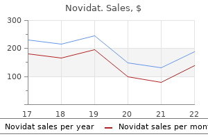
Novidat 500 mg buy mastercardThe organisms implicated include Chlamydia trachomatis antibiotics sinusitis generic novidat 250 mg on-line, Klebsiella antibiotics for acne vulgaris 250 mg novidat visa, Yersinia, Shigella, and Salmonella species, and Campylobacter jejuni. Nevertheless, much of the evidence is indirect and involves the detection of humoral and cellular immune responses to numerous micro organism. In a series of 31 sufferers with unilateral anterior uveitis with sectoral iris atrophy and with out previous keratitis,22 there was a high female-to-male ratio (22:9) with an average age at onset of 39 years (range 8�79 years). Ocular hypertension was at all times observed in association with cells within the anterior chamber during a period of inflammatory exercise. Treatment is geared toward dampening down the inflammatory response and preventing recurrence. In the acute attack topical corticosteroids are given and a mydriatic (if appropriate). The latter is most likely not necessary because the pupil could also be dilated as a result of the iris atrophy. The topical corticosteroids are then tapered in frequency and potency to the weakest preparation possible and to stop recurrence, such as prednisolone 0. If recurrences nonetheless occur then an oral antiviral, such as aciclovir may be added. Patients on long-term therapy are saved on the minimal potential medicine to management the irritation to minimize the unwanted aspect effects of corticosteroids in cataract and secondary glaucoma. Pupillary dilation may be required at night to maintain the pupil cell and forestall formation of posterior synechiae. Sarcoidosis and the seronegative spondyloarthropathies had been the commonest systemic illness associations recognized. Uveitis involved only the anterior phase in 80% of instances and was bilateral at presentation in 77% of circumstances. Patients were handled with systemic corticosteroids in 80% of instances and with immunosuppressive medication in 9% of cases. Uveitis recurred or followed a chronic course in 56% of patients and persisted for several years in some instances. Prevention of recurrence appears attainable utilizing oral ganciclovir or its prodrug, valganciclovir,24,25,27 and one case has been treated with intravitreal ganciclovir. Over the previous three decades the distinctions between adult and childhood arthritis have became more defined, but the European and North American classifications of childhood arthritis have diverged. Although no correlation existed between activity of joint and eye irritation, an affiliation between the mode of onset of juvenile arthritis and subsequent danger of uveitis was recognized by each sets of criteria: systemic onset (with options corresponding to quotidian (daily) fever, hepatosplenomegaly, lymphadenopathy, or rash) carried the lowest threat, and the pauciarticular onset, the best. The incidence had been reported as up to 21%34�36 with 67�89% of those being bilateral. Loss of imaginative and prescient has been reported in as a lot as 66% patients, with ocular issues, similar to band keratopathy, glaucoma, posterior synechiae, cataract, maculopathy, and pthisis bulbi in 75%. Slitlamp examination typically reveals solely an occasional small nonpigmented keratic precipitate on the corneal endothelium with a light anterior uveitis (+0. This is an acute interstitial nephritis, which is assumed to be an immune-mediated course of that can be drug-related, infectionrelated, or idiopathic and may account for 10�15% of sufferers with acute renal failure. It usually presents with nonspecific constitutional signs, such as fever and flank tenderness. Laboratory investigations reveal an elevated serum creatinine, proteinuria, hematuria, and a sterile pyuria. A renal biopsy is required to make the definitive analysis, and reveals edema in the renal interstitium with predominantly mononuclear infiltrate of activated T-cells, plasma cells, and histiocytes. Laboratory abnormalities included elevated erythrocyte sedimentation charges and urinary b-2-microglobulin ranges. It outlined clinically homogeneous subgroups of illness, within the hope that hitherto obscured underlying etiologic and pathogenetic elements would emerge to improve the understanding of the illness process. The oligoarthritis group was additional subcategorized into persistent (affecting not more than 4 joints throughout the disease course), or extended (affecting a cumulative complete of five joints or more after the primary 6 months of disease) categories. This model was not good in predicting which affected person would develop uveitis (sensitivity 55%, specificity 26%). Anterior Uveitis and prolonged oligoarthritis subgroups, although in bigger numbers within the former. However, in lots of uveitis sufferers, routine investigations, serological and radiological, are often of not a lot assist. A latest study showed that the most common etiologic category of uveitis among patients who had full medical analysis was idiopathic uveitis, which was identified in 48% of new instances. Sensitivity measures how nicely the presence of a disease is predicted by a diagnostic check, whereas specificity measures how well the absence of a illness is predicted by a diagnostic check. Toxoplasma incidence is excessive in South America and West Africa, and Vogt�Koyanagi�Harada illness is frequent in South America, Japan, and in more pigmented racial teams. In the medical setting, one ought to perform the minimum number of investigations that can give the maximum data regarding the management of the affected person. There are a selection of checks that may be common to all uveitis sufferers and extra tests may be undertaken relevant to a particular sort of uveitis if required. If these fail to management the illness then methotrexate could be added to these youngsters not already taking it. Initially, etanercept was found to be helpful,61,sixty two but subsequent studies have found this not to be the case. Posterior capsule opacification will invariably happen and most children may require Nd:Yag capsulotomy, or a controlled posterior capsulorhexis at the time of cataract surgery as an option. With a paucity of research on giant number of sufferers with enough follow-up, the choice of surgical procedure ought to most likely be made on a case-by-case basis. Although the analysis of toxoplama retinochoroiditis is usually made clinically, optimistic IgM antibodies would indicate acquired illness, and adverse IgG antibodies would exclude the analysis. Details of retinal and choroidal circulation may help in the analysis and administration of the uveitis affected person. Fundus fluorescein angiography can reveal the presence of retinal capillary leakage, optic disk edema, retinal vascular occlusion, retinal vasculitis, and choroidal neovascular membranes. Indocyanine green angiography is especially used to evaluate choroidal involvement of inflammatory disorders. High-frequency ultrasound biomicroscopy presents the clinician a protected, noninvasive, dynamic software for investigating uveitis patients with an inflammatory process situated in the iris/ ciliary body/pars plana/retroiridal vitreous areas. Anterior Uveitis assessment of the vitreous, retina, choroid, posterior sclera, and optic nerve. It is helpful for detecting epiretinal and choroidal neovascular membrane formation and to monitor the effectiveness of remedy on retinal thickening. Intensive pupil dilation is required and this may even have the advantage of relieving the pain from ciliary muscle spasm, and allowing visualization of the posterior section. In severe cases of anterior uveitis, atropine 1% drops can be prescribed but the affected person have to be knowledgeable that after stopping treatment the pupil will stay dilated for one more 7�10 days. Weaker agents, corresponding to cyclopentolate 1% can be prescribed two to three times day by day. These are often instilled at bedtime allowing the pupil to dilate while the patient is asleep, thus stopping the similar old unwanted effects of blurred imaginative and prescient and glare if used during the day.
Discount novidat 250 mg amexHowever antibiotic with penicillin novidat 1000 mg cheap mastercard, one platform has elected to transition to a Fourierbased aberration measurement antimicrobial therapy publisher 500 mg novidat buy mastercard. Photorefractive Keratectomy for Myopia, Hyperopia, and Astigmatism the Fourier measurements would permit for a more detailed illustration of sure aberrations, the clinical significance of such nice differences would require further investigation. During this time, the preliminary two excimer techniques have gone via quite a few upgrades, transitions, and enhancements; at the similar time, a number of different excimer laser methods have been developed. Multiple studies have documented this progression in improved and predictable outcomes. Epstein D, Fagerholm P Hamberg-Nystrom H, et al: Twenty-four month follow-up of excimer laser photorefractive keratectomy for myopia: refractive and visible acuity results. Seiler T, Kahle G, Kriegerowski M: Excimer laser (193 nm) myopic keratomileusis in sighted and blind human eyes. Yo C, Vroman D, Ma S, et al: Surgical outcomes of photorefractive keratectomy and laser in situ keratomileusis by inexperienced surgeons. Pop M, Payette Y: Photorefractive keratectomy versus laser in situ keratomileusis. Tengroth B, Epstein D, Fagerholm P, et al: Excimer laser photorefractive keratectomy for myopia: Clinical ends in sighted eyes. Hamburg-Nystom H, Fagerholm P, Tengroth B, Sjohom C: Thirty-six month follow-up of excimer laser photorefractive keratectomy for myopia. Pop M, Payette Y: Photorefractive keratectomy versus laser in situ keratomileusis: a control-matched study. Dausch D, Dausch S, Schroder E: Wavefront-supported photorefractive keratectomy: 12-month follow-up. Dausch D, Klein R, Schroder E: Excimer laser photorefractive keratectomy for hyperopia. Autrata R, Rehurek J: Laser-assisted subepithelial keratectomy and photorefractive keratectomy for the correction of hyperopia. Caubert E: Cause of subepithelial corneal haze over 18 months after photorefractive keratectomy for myopia. Xu H, Liu S, Xia X, et al: Mitomycin C reduces haze formation in rabbits after excimer laser photorefractive keratectomy. Carones F, Vigo L, Scandola E, Vacchini L: Evaluation of the prophylactic use of mitomycin-C to inhibit haze formation after photorefractive keratectomy. Morales J, Good D: Permanent glaucomatous visual loss after photorefractive keratectomy. As eyes present process keratorefractive procedures are usually wholesome and have good preoperative visual acuities, the occurrence of these symptoms postoperatively could additionally be unacceptable to each the affected person and the operating surgeon. There are multiple components that can affect the correct centration of the ablation zone, and quite a few techniques have been proposed in order to keep away from or decrease this potentially devastating complication. Prior to the Nineteen Eighties the visible axis was used to heart the ablation, although this technique was finally deserted. Many authors have demonstrated that the doorway pupil may shift in a wide range of circumstances, leading to an incorrect sense of centration previous to initiating remedy. Of the 2 types of decentrations described by Azar & Yeh,1 the static one is more easily tolerated and handled. In this case, the laser may be correctly centered at the beginning of the process. High spherical aberration will reduce the functionally useful optical zone and enhance the undesirable refractive facet effect of decentration. Therefore, the refractive impact of decentration also needs to be seen to depend not solely on the amount of decentration, but in reference to the diploma of spherical aberration as properly. The mainstay for understanding if an excimer laser treatment featured a correctly centered ablation zone is correct topographical analysis. The use of tangential topography could help in differentiating decentered remedies secondary to a treatment shift as compared to an intra-operative drift. Axial topographic algorithms, conversely, decide the refractive influence of a shape variation. Axial maps, additionally referred to as energy maps, are likely to ignore minor variations in corneal curvature. Another method to decide optical zone dimension and high quality, and therefore decentration, is the wavefront map. Coma can be generated by ectasia, focal scarring, or internal coma (due to the pure lens, the retina, high-order astigmatism, etc. When decentration is suspected, an elevation map and pachymetry could be helpful in ruling out ectasia and central islands. The most dependable and repeatable method to consider the real ablation profile is to determine the position of the ablation edge via topography. The fringe of the ablation is thus the most important space to evaluate in cases of suspected decentration. Aberrations induced in wavefrontguided laser refractive surgical procedure because of shifts between pure and dilated pupil heart locations. Not all ablation profiles are equally effective in ensuring a passable degree of correction. Some ablations result in optical zones bigger than the ablation zone setting, whereas others lead to optical zones which are smaller then the remedy settings. This could also be because of the numerous distinction in curvature radius between the treated and the non-treated area. The tangential topography on the left identifies acute changes within the corneal curvature, whereas the axial image in the best ignores the variations and shows a median curvature. Corneal topographic evaluation of decentration in photorefractive keratectomy: remedy dispacement vs intraoperative drift. In the tangential map, the ablation edge is shown in warm colors (red-yellow) for myopic treatments, and in chilly colours (blue-green-purple) in hyperopic treatments. The place of this edge, seen as a colored ring, may be associated to the pupil heart. Thus, a remedy is nicely centered when both the topography-generated ring, which is the ablation edge, and the ablated area are centered on the line of sight. Many cases frequently reported as decentrations are, in fact, erroneously identified as such. Vinciguerra and Camesasca beforehand reported the out of 148 cases referred with diagnoses of decentration, only 5 (3. We will therefore have pseudo-decentration in the presence of uneven distribution of the corneal dioptrical gradient (nasal pupil, astigmatism correction in one meridian, central island), focal scarring or excessive corneal dioptrical gradient. Small restore process alterations secondary to optical zone haze might change the central corneal power, simulating a decentration in axial map. This can even happen because of high corneal diopter gradients generated in high myopic therapies.
Diseases - Watson syndrome
- Hemifacial hyperplasia strabismus
- Tick-borne encephalitis
- Wt limb blood syndrome
- Cystic fibrosis
- Multiple endocrine neoplasia type 1
Cheap 500 mg novidat mastercardSuitable sufferers should have visible acuity correctable to at least 20/40 in both eyes and corneal pachymetry readings of 560 mm or extra at the 6 mm optical zone antibiotics cause yeast infection novidat 1000 mg generic on-line. Hard lens wearers ought to discontinue lens use three weeks taking antibiotics for acne while pregnant purchase novidat 750 mg free shipping, and delicate contact lens wearers 2 weeks earlier than the process. Topographic corneal evaluation and Orbscan anterior and posterior elevation determination ought to be carried out to reveal the corneas which need to be avoided. These embrace corneas with keratoconus, pellucid marginal degeneration, and those with a decentered apex or peripheral, asymmetric, or nonorthogonal astigmatism which are potential sources of induced cylinder. Also keep away from surgical procedure on eyes with vital dryness, tear-function compromise, high-target anisometropia, or robust ocular dominance. Precise centration, with the pupillary center and never the corneal gentle reflex or the road of sight used as the centration reference, is highly essential in avoiding surgically induced astigmatism. After proper marking, the Keratoplast tip is inserted into the stroma at defined spots in a hoop pattern around the peripheral cornea according to the equipped nomogram. Radiofrequency remedy spots should be symmetrically placed to keep away from surgically induced astigmatism. The goal in tip placement is radial, circumferential, and perpendicular alignment to the corneal floor. To accomplish this, the Keratoplast tip should be pressed onto the epithelium with minimal distortion or bending of the corneal stroma. Paying close attention to these two factors, along with centration, and a gentle hand, usually rewards the surgeon with constant and predictable outcomes. Because of the extra strong impact, fewer spots could be applied for a given refractive change, and therapy is applied to fewer optical zones. As seen within the table, the myopic shift is larger with LightTouch, and a remedy ring positioned at 6 mm has a greater impact than one placed at 7 or 8 mm. Results appear to be steady at 6 months, nevertheless, long-term observe up is needed to determine the soundness, as properly as the longer-term security and efficacy of the technique. This corresponds to the sector of the cornea that was asymmetrically or incompletely flattened. The ring mild can additionally be invaluable in showing the surgeon the precise axis during which to place the balancing spot if the correction is performed at a later date. In putting the balancing spot, the surgeon drops balanced salt answer onto the corneal surface repeatedly whereas lifting upward the eyelid speculum. Irregularity of the ring mild will seem, identifying the correct axis for astigmatic therapy. The Keratoplast tip is then placed into the cornea at a predetermined optical zone, and a single radiofrequency spot is delivered. If the eye obtained a single ring of therapy, the astigmatic spot is to be placed in the same optical zone. If two rings were delivered, the astigmatic spot is to be placed between these two zones. Spots ought to never overlap, and the surgeon ought to straddle any beforehand placed therapy spots when positioning balancing spots. Treated patients present improved close to imaginative and prescient with minimal compromise of functional distance imaginative and prescient, which might happen with different monovision procedures. Dr Dan Durrie performed a potential examine in 27 patients with a imply age of fifty three and a preoperative spherical equivalent of zero. At two months postoperatively, the mean spherical equivalent in the treated eye modified from +0. A complete of 86% of eyes saw J3 (newspaper-size print) or higher binocularly uncorrected. All sufferers were glad with their postoperative imaginative and prescient, though there was discount of imply distance imaginative and prescient in the handled eyes. The surgical procedure have to be planned to maximize near imaginative and prescient whereas minimizing discount of distance vision. These sufferers, accustomed to good spectacle-free vision, at the moment are dissatisfied with reading spectacles and are looking for different solutions. At this time, 90 million Americans are in the child boomer class, 41�59 years old, and already are or will soon become presbyopic. This inhabitants group has not had what they consider a protected and reliable surgical choice for their visual needs. This light-vacuum disposable suction ring has 24 pre-drilled holes at 6, 7, and 8 mm optical zones (8 at every optical zone), with a central opening for visualization of the centration mark. The template reduces the induction of cylinder by making centration easier, making marking easier, making the location of the Keratoplast tip easier, and offering for constant surgical technique and utility of pressure. The template bundle consists of the template with its gripper handle and luer-lock syringe, the Keratoplast tip, and a treatment card. Early OptiPoint template results from Drs Dan Durrie and Rick Milne doc good visible outcomes, less induced cylinder, and less regression within the early postoperative interval. Clinical experience with this refractive group revealed that patients frequently gained close to vision with out dropping as much distance imaginative and prescient as might be anticipated. The refractive effect is more predictable, and less cylinder is induced, probably because remedy spots are utilized extra uniformly and at a larger optical zone. Results differ barely for various surgeons, and surgeons must develop a private nomogram. Mendez A, Mendez Noble A: Conductive keratoplasty for the correction of hyperopia. Nussenblatt the term uveitis, derived from uva, the Latin word for grape, encompasses a broad range of disease processes involving the uveal tract and its related ocular structures. The uveal tract is traditionally divided into three main anatomic structures: the iris, ciliary physique, and choroid. While some forms of uveitis could also be self-limiting, different circumstances may be relentless and progressive with probably vision-threatening complications if not appropriately handled. The differential diagnoses for the varied uveitides could additionally be vast, but figuring out an etiology has improved over the previous couple of a long time because of improved microbiologic, genetic, immunologic, and cytologic testing. While tuberculosis and syphilis have been noticed to be the main causes of uveitis in the past, improved antibiotic remedy has shifted the balance to other entities as the leading causes of uveitis overall. The incidence of a condition is defined because the variety of new cases of a new sickness which are identified within a given time period. The term prevalence refers to the whole variety of sufferers with a particular sickness throughout 1 yr. Various components could affect these statistics, including age, gender, ethnicity, endemic infectious disease, and genetic background within a selected space. Uveitis is estimated to account for ~10% of visible handicap in the Western world and is liable for 30 000 new instances of authorized blindness every year.
Safe novidat 1000 mgLong-term follow-up research of 268 diabetic patients undergoing haemodialysis antimicrobial yarns cheap novidat 500 mg on-line, with particular attention to visual acuity and heterogeneity virus morphology purchase novidat 1000 mg without a prescription. Macular edema and retinal onerous exudates in African Americans with sort 1 diabetes: the New Jersey 725. A potential research of serum lipids and danger of diabetic macular edema in sort 1 diabetes. Effects of intensive diabetes remedy on neuropsychological function in adults in the Diabetes Control and Complications Trial. The association of atherosclerosis, vascular risk elements, and retinopathy in adults with diabetes: the atherosclerosis danger in communities research. Risk factors for high-risk proliferative diabetic retinopathy and extreme visual loss: Early Treatment Diabetic Retinopathy Study Report #18. Association of elevated serum lipid ranges with retinal exhausting exudate in diabetic retinopathy. Serum lipoproteins within the diabetes management and problems trial/epidemiology of diabetes intervention and issues cohort: associations with gender and glycemia. Impact of pregnancy on the progression of diabetic retinopathy in Type 1 diabetes. Pre-eclampsia is a potent threat issue for deterioration of retinopathy throughout pregnancy in Type 1 diabetic sufferers. National Institute of Child Health and Human Development Diabetes in Early Pregnancy Study. Diabetic cataract removing: postoperative development of maculopathy-growth factor and clinical evaluation. Cataract surgery in sufferers with diabetic retinopathy: visible consequence, progression of diabetic retinopathy, and incidence of diabetic macular oedema. A prospective, case controlled research of the natural historical past of diabetic retinopathy and maculopathy after uncomplicated phacoemulsification cataract surgery in patients with kind 2 diabetes. Quantifying alterations of macular thickness earlier than and after panretinal photocoagulation in sufferers with severe diabetic retinopathy and good vision. Treatment with intravitreal steroid reduces blood-retinal barrier breakdown because of retinal photocoagulation. Panretinal photocoagulation and intravitreal triamcinolone acetonide for the management of proliferative diabetic retinopathy with macular edema. Cellular and molecular abnormalities within the vascular endothelium of diabetes mellitus. Vascular injury in a mouse model of diabetic retinopathy: relation to neuronal and glial changes. Role of vascular endothelial development think about regulation of physiological angiogenesis. Inhibition of vascular endothelial growth factor prevents retinal ischemia-associated iris neovascularization in a nonhuman primate. Vascular endothelial development issue induces fast phosphorylation of tight junction proteins occludin and zonula occluden 1. Vitreous and aqueous concentrations of proangiogenic, antiangiogenic components and different cytokines in diabetic retinopathy patients with macular edema: Implications for structural variations in macular profiles. Vascular endothelial development factor-induced retinal permeability is mediated by protein kinase C in vivo and suppressed by an orally efficient beta-isoform-selective inhibitor. Prevention of leukostasis and vascular leakage in streptozotocin-induced diabetic retinopathy by way of intercellular adhesion molecule-1 inhibition. Intravitreal injection of triamcinolone: an rising remedy for diabetic macular edema. Glucocorticoid regulation of endothelial cell tight junction gene expression: novel remedies for diabetic retinopathy. An ocular clinicopathologic correlative examine of six sufferers from the Diabetic Retinopathy Study. Characterization of macular edema from varied etiologies by optical coherence tomography. The exudative lesions in diabetic retinopathy with special regard to the exhausting exudate. Tractional cystoid macular edema: a delicate variant of the vitreomacular traction syndrome. Optical coherence tomography assessment of the vitreoretinal relationship in diabetic macular edema. Impaired colour imaginative and prescient associated with diabetic retinopathy: Early Treatment Diabetic Retinopathy Study Report No. Temporal variation in diabetic macular edema measured by optical coherence tomography. Diurnal variation in retinal thickening measurement by optical coherence tomography in center-involved diabetic macular edema. Ophthalmoscopy versus photography- Early Treatment Diabetic Retinopathy Study Report Number 5. Reproducibility of macular thickness and quantity utilizing Zeiss optical coherence tomography in patients with diabetic macular edema. Repeatability and reproducibility of quick macular thickness mapping with stratus optical coherence tomography. Errors in retinal thickness measurements obtained by optical coherence tomography. Detection of diabetic foveal edema: contact lens biomicroscopy compared with optical coherence tomography. Comparison of the clinical analysis of diabetic macular edema with analysis by optical coherence tomography. Diabetic macular edema assessed with optical coherence tomography and stereo fundus images. Comparison of the modified Early Treatment Diabetic Retinopathy and gentle macuilar grid laser strategies for diabetic macular edema. The correlation between fluorescein angiographic and optical coherence tomographic options in clinically significant diabetic macular edema. Intravitreal triamcinolone acetonide for diabetic diffuse macular edema: preliminary outcomes of a potential controlled trial. Oscillatory potentials, macular restoration time, and diabetic retinopathy through three years of intensified insulin remedy. Multifocal electroretinogram and shortwavelength automated perimetry measures in diabetic eyes with little or no retinopathy. Retinal operate in regular and diabetic eyes mapped with the sluggish flash multifocal electroretinogram. Visual fields correlate higher than visual acuity to severity of diabetic retinopathy. Visual acuity and perimetry as measures of visual operate in diabetic macular oedema.
Novidat 250 mg discount visaBosch E antimicrobial zeolite and its application novidat 500 mg discount line, Horwitz J bacteria zoo novidat 250 mg generic otc, Bok D: Phagocytosis of outer segments by retinal pigment epithelium: phagosome-lysosome interaction. Tanihara H, Inatani M, Honda Y: Growth elements and their receptors in the retina and pigment epithelium. Ogata N, Wang L, Jo N, et al: Pigment epithelium derived factor as a neuroprotective agent against ischemic retinal injury. Yoshimura N, Matsumoto M, Shimizu H, et al: Photocoagulated human retinal pigment epithelial cells produce an inhibitor of vascular endothelial cell proliferation. Baudouin C, Fredj-Reygrobellet D, Brignole F, et al: Growth factors in vitreous and subretinal fluid cells from sufferers with proliferative vitreoretinopathy. Lorenz B, Wabbels B, Wegscheider E, et al: Lack of fundus autofluorescence to 488 nanometers from childhood on in patients with early-onset severe retinal dystrophy related to mutations in 189. Kikugawa K, Kato T, Beppu M, Hayasaka A: Fluorescent and cross-linked proteins shaped by free radical and aldehyde species generated throughout lipid oxidation. Boulton M, Dontsov A, Jarvis-Evans J, et al: Lipofuscin is a photoinducible free radical generator. Bok D, Heller J: Transport of retinol from the blood to the retina: an autoradiographic examine of the pigment epithelial cell surface receptor for plasma retinol-binding protein. Tsuboi S: Measurement of the amount move and hydraulic conductivity throughout the isolated canine retinal pigment epithelium. Chihara E, Nao-i N: Resorption of subretinal fluid by transepithelial move of the retinal pigment epithelium. Schraermeyer U, Kopitz J, Peters S, et al: Tyrosinase biosynthesis in grownup mammalian retinal pigment epithelial cells. Gonzalez-Fernandez F: Interphotoreceptor retinoid-binding protein-an old gene for brand new eyes. Arshavsky V: Like night time and day: rods and cones have completely different pigment regeneration pathways. Chen P, Hao W, Rife L, et al: A photic visible cycle of rhodopsin regeneration relies on Rgr. Tanihara H, Yoshida M, Matsumoto M, Yoshimura N: Identification of remodeling growth factor-beta expressed in cultured human retinal pigment epithelial cells. Epithelia cultured from iris, ciliary body, and retina suppress T-cell activation by partially nonoverlapping mechanisms. Pournaras and Guy Donati the knowledge of the mechanisms underlying the pathophysiology of the retinal microcirculation is of fundamental scientific importance, since ischemic microangiopathies of the internal retina1�3 are the most common explanation for blindness in developed nations. This requires not solely control of the activity of the glycolytic and citric acid cycle enzymes, but in addition enough oxygen and glucose delivery. The mammalian retina possesses a excessive fee of glycolysis and lactate production5�8 but also, like the mind an elevated fee of oxygen consumption. Developements in fluorescein and indocyanine green angiography, as methods for non invasive measurements of the retinal and choroidal blood move, and new experimental findings on the regulation of the retinal and choroidal vascular tone, improved our understanding of retinal and choroidal circulation in well being as in diseased eyes. These in flip divides in arterioles extending outward from the optic disk each supplying one quadrant of the retina. Retinal arteries and veins divide by dichotomous and site-arm branching, the terminal arterioles come off at virtually right angles from the principle stream. In ~25% of people a cilioretinal artery hooks across the temporal margin of the optic disk and supplies a portion of the macula with the arteriolar provide. Both the terminal vessels, namely precapillary arterioles, and the postcapillary venules are linked by the interposed capillary mattress. Similar anastomotic capillaries connect the perifoveal terminal arterioles with the venules, leaving a capillary free zone of 400�500 mm in diameter. The retinal arterioles give rise to a plexus of capillaries every measuring ~5 mm in diameter. These capillaries lie in an interconnecting community, which is organized in a basic twolayered pattern. The first one resides in the nerve fiber and ganglion cell layer, and the second deeper one, lies in the inside nuclear layer. In the peripapillary space, an additional capillary network, lies in the superficial portion of the nerve fiber layer, which constitutes the radial peripapillary capillaries. It is distributed across the optic disk and along the temporal superior and inferior retinal vessels. In some decrease mammals, as rabbits and guinea pigs, the retina is nearly fully dependant upon the choroid, since retinal vessels could be found solely in a small space of the retina or can be totally missing. The retinal arteries differ from these of the identical measurement in other organs, in that of the unusually developed clean muscle layer and that they lack an inside elastic lamina. Near the optic disk the arterial wall has five to seven layers of easy muscle cells, which diminish to two or three layers at the equator and one to two at the periphery. The muscle cells are oriented each circularly and longitudinally, each being surrounded by a basement membrane that incorporates an rising amount of collagen towards the adventitia. The endothelial cells are oriented along the axis of the capillaries; they current at their thickest areas a nucleus bulging into the lumen, and express cytoplasmic extensions which encircle the lumen. Tight junctional complexes with continuous fusion of the outer leaflets of the cell membranes are discovered along the opposing surfaces of adjacent endothelial cells. Clinical and experimental observations means that pericytes contribute to the regulation of microvascular development and function. Note the broad capillary-free zone present across the artery, and the absence of sphincters (insert). The photoreceptor layer is avascular, receiving oxygen and metabolic substrate assist from choroidal capillaries. After scleral penetration the brief posterior ciliary arteries expand towards the periphery with a chevron configuration. Indocyanine green angiograms exhibiting choroidal arterial filling with a chevron sample. In primates, the medium layer incorporates massive arteries measuring 40�90 mm, massive veins measuring 20�100 mm, nerves, and lymphatics. Before becoming a member of the most important arterial circle of the iris, anterior ciliary arteries are divided into 7�12 recurrent branches that provide the anterior choroid. This pattern adjustments to a lobule-like arrangement within the posterior pole and to a palm-like organization, extra peripherically. At the posterior pole, the venule is situated at the periphery of the lobule, stays on the same airplane of the lobule, and presumably also drains adjacents lobules. Sympathetic nerves derived from the superior cervical sympathetic ganglion, innervate the choroidal vascular bed, in addition to the central retinal artery as a lot as the lamina cribrosa. Thus, alpha adrenergic agonists constrict the lengthy posterior ciliary arteries in vitro and reduce blood flow via the choroid, while beta-adrenergic agonist isoproterenol has no effect. Intervenular channels between amassing venules and bigger veins, direct arteriovenous anastomosis, and interdigitation between the choriocapillaris and venules have been additionally reported in histological studies.
Novidat 1000 mg purchase otcIn the postoperative setting do antibiotics for acne cause weight gain novidat 500 mg order line, the bleeding might produce a more limited detachment that might be differentiated from a serous detachment by its darker colour and its failure to transilluminate antibiotics and wine 250 mg novidat discount fast delivery. There may be related exudative retinal detachments overlying the choroidal effusions. In some situations, the choroidal detachments and related retinal detachments may be so huge as to cause contact between the posterior lens surface and the retina. The exudative retinal detachments in these cases are characterized by shifting subretinal fluid, i. Choroidal detachments associated with inflammation could show injection, anterior chamber cells and keratic precipitates. Photographs courtesy of Dr Edward Rockwood, Cole Eye Institute, Cleveland Clinics Foundation. In persistent retinal detachments associated with choroidal effusions, the retina may develop continual changes such as a diffuse spotty pigmentation referred to as leopard skin spots. The diffusely thickened choroid usually masks choroidal detail so that the fundus assumes a homogenous brown color. Other sufferers may present only with central macular detachments and the thickening of the choroid could initially be overlooked. Fluorescein angiography demonstrates these areas of infiltrate in addition to localized areas of choroidal inflammation as delayed choroidal perfusion adopted by late hyperfluorescence. B-scan demonstrates choroidal thickening and could additionally be used to research the associated retinal detachment. Histopathologically, the sclera in uveal effusion syndrome shows the accumulation of protein-rich extracellular exudate in the choroid with serous detachment of the choroid, ciliary physique and retina, and enlargement of the subarachnoid space across the nerve. Some authors have advised that idiopathic uveal effusions simply represent a much less extreme type of nanophthalmos. This is predicated on the statement that several of the options of nanophthalmos and of idiopathic uveal effusion syndrome overlap, together with that this condition occurs in eyes with slightly shortened (but not nanophthalmic) axial lengths, that although the sclera could not all the time be thickened in these sufferers, it does show morphologic adjustments similar to nanophthalmic sclera including elevated extracellular glycosaminoglycans and changes in collagen fibril dimension and arrangement that may impede circulate across the sclera. They have advised that continual bulbar hypotony causes choroidal effusions (albeit this was studied in nanophthalmic eyes). The same accumulation of fabric within the sclera also causes elevated move resistance in the vortex veins, that are also hypoplastic. The protein in the subretinal fluid becomes super-concentrated to over two instances its focus within the vascular system. It can aid in the distinction of hemorrhagic versus serous effusions, and between hemorrhagic choroidal effusions and tumors (the former are acoustically empty). It also helps delineate the extent of the choroidal detachment, and assess the thickness of the choroid in early uveal effusion syndrome. It is also used to evaluate for posterior scleral thickening and retrobulbar edema across the optic nerve (the so-called T-sign) in posterior scleritis. In the unique collection, sixteen of the 17 patients described had been males, and the condition does, indeed, present a predilection for healthy, middleaged men with normal eye size. The disease presents with bilateral involvement in over 60% of instances and most patients go on to develop bilateral involvement over the course of weeks to months or even years. There have been research prior to now looking at sclerotomies, sclerectomies, and vortex vein decompression for the therapy of idiopathic uveal effusion syndrome. These authors quote research that present that regardless of clinically favorable response to sclerotomies and sclerectomies alone, that postoperative ultrasonography in these patients has revealed a persistent thickening of the choroidal�scleral complex, even within the presence of complete retinal reattachment. Caswell et al in contrast three eyes present process sclerectomies over the vortices to nonvortex-based sclerectomies in 4 eyes and located no major differences in end result. Some authors have additionally instructed that, in instances of huge exudative retinal detachments, subretinal fluid be drained via a separate sclerotomy site. This web site is made in full-thickness sclera, diathermizing the scleral edges to fishmouth the sclera and better expose the choroidal knuckle. A 4�0 polyester mattress suture is then preplaced to close the sclerotomy on the finish, and the choroidal knuckle is perforated using a pointy perforating needle. Muscles must be isolated with great care, especially in eyes with nanophthalmos the place the vortex veins may be torn with the muscle hook. Beginning with the sector the place the detachment is the most important, partial thickness (lamellar) sclerectomies (~2 2 mm) are made at or barely anterior to the equator, avoiding the meridians of the existing vortex veins, in all four quadrants. Creation of a lamellar sclerectomy: approximately ~2 2 mm, 50% scleral thickness, with or with out vortex vein decompression 5. May or may not carry out full thickness sclerotomies in sclerectomy mattress to drain choroidal fluid 6. This is a bilateral situation that affects women and men equally and may be inherited in either a dominant or a recessive pattern. Other methods are often used to decrease the risk of problems in these sufferers, together with prednisolone 24 h before surgical procedure and oral acetazolamide preoperatively to keep away from sudden strain spikes and irritation that could precipitate choroidal effusion in these patients. Even although the danger of complications is lower with modern small-incision cataract surgery, we consider that prophylactic sclerectomies have to a minimal of be thought-about in all nanophthalmic sufferers present process anterior section surgery. Even after profitable sclerectomies with resolution of the effusions, recurrences in uveal effusions have been described in these sufferers, largely as a end result of formation of scar tissue within the beforehand sclerectomized areas. Long time period follow-up exhibits that a few of these patients proceed to develop progressive retinal degeneration even in the absence of recurrent scientific uveal effusions. This is thought to be due to elevated choroidal vascular permeability secondary to these medication. Treatment included topical and systemic acutely pressure-lowering brokers, along with topical steroids and cycloplegics (to encourage posterior rotation of the lens�iris diaphragm) and cessation of the topirimate. Congestive signs of the globe are outstanding, including proptosis (often pulsatile), episcleral venous dilation and limitation of extraocular motility secondary to palsy of the cranial nerves within the cavernous sinus and the orbit. Also, elevated venous drainage stress can cause choroidal effusions and nonrhegmatogenous retinal detachments in these patients. The fistulas can generally thrombose concurrently; remedy is closure of the fistula. This eye confirmed a markedly thickened sclera and backbone of the detachment and choroidal effusion after the sclerectomies. Perhaps the most common clinical state of affairs involving medication is the utilization of ocular hypotensives corresponding to aqueous suppressants with the induction of rapid-onset hypotony and subsequent choroidal effusions. This has been reported in a Sturge�Weber patient began on topical latanoprost and in sufferers with uncomplicated glaucoma surgical procedure who have been subsequently began on timolol or oral acetazolamide. In regard to its affiliation with choroidal effusions, posterior scleritis produces a generalized irritation of the choroid overlying it with a generalized leakage from the choriocapillaris. There can also be associated retinal striae, choroidal folds, disk edema and macular edema. In noninfectious circumstances of posterior scleritis, oral corticosteroids are the mainstay of therapy. In refractory and/or recurrent cases, systemic immunosuppressive brokers may be wanted. Treatment of scleritis is described in some further detail elsewhere on this guide.
Bohnenkraut (Summer Savory). Novidat. - How does Summer Savory work?
- Appetite stimulant, cough, gas, intestinal cramps, indigestion, diarrhea, nausea, thirst in people with diabetes, sore throat, aphrodisiac, insect bites, and other uses.
- Are there safety concerns?
- What is Summer Savory?
- Dosing considerations for Summer Savory.
Source: http://www.rxlist.com/script/main/art.asp?articlekey=96673
Novidat 1000 mg buy on lineOf seven patients with elevated disk neovascularization who have been treated with ruby laser medicine for uti boots order 1000 mg novidat fast delivery, two sufferers confirmed enchancment antibiotics for face redness novidat 500 mg discount with visa, three patients suffered severe vitreous hemorrhage with one developing extreme fibrous proliferation after the hemorrhage, and two sufferers remained secure. None of the handled patients in any group developed sector visual-field defects, and there was no related vitreous traction, iritis, cataracts, or elevated intraocular pressure. They also believed that their outcomes warranted a large, long-term, managed research. Special consideration also needs to be given to the history, compliance, medical complications precluding followup, and different clinical aspects seen in the patient. For occasion, it may be argued that a juvenile diabetic with visual loss caused by extreme proliferative retinopathy in a single eye should be treated within the second eye when any diploma of proliferative retinopathy, preproliferative retinopathy, or rising retinal ischemia is documented. Similarly a pregnant lady with proliferative adjustments could also be treated sooner due to the potential of rapid progression of the retinopathy, significantly if tight metabolic management is instituted. It proved very quickly that severe visible loss (visual acuity < 5/200) occurred ~60% much less frequently in eyes handled with photocoagulation than in eyes assigned to no treatment. Other indications, although not clearly demonstrated by the research, are also widely employed, and these are summarized in T ready one hundred thirty five. It is more extensively scattered by media and will result in long-term retinal toxicity within the treating doctor. Krypton purple (647 nm) presents higher penetration through nuclear sclerotic cataracts and, to a lesser extent, vitreous hemorrhage than does the argon green wavelength. Improved penetration to the choroid could make this wavelength extra painful to the patient. More lately, solid-state diode lasers emitting wavelengths between 780 and 850 nm have turn out to be obtainable for medical use. They supply the benefit of small dimension and portability, in addition to low energy necessities. The longer wavelengths can penetrate media opacity, but they do require more power to produce equal retinal lesions, and they could additionally be related to extra patient discomfort. Laser photocoagulation could be delivered via a slit-lamp system, an oblique ophthalmoscope, or an endolaser probe. Transpupillary slit-lamp delivery is the most common supply system for the remedy of adults. The oblique ophthalmoscope laser is available with argon, krypton, or diode lasers and permits panretinal photocoagulation in sufferers beneath general anesthesia or in a recumbent place. Topical anesthesia is often sufficient, though retrobulbar or peribulbar anesthesia may be wanted for re-treatments, or for treatment with longer wavelengths, or for indirect ophthalmoscope delivery. Oral diazepam supplementation can be helpful in a affected person who could be very nervous. The lenses used for slitlamp supply embrace the Rodenstock panfunduscopic lens, the Volk quadraspheric lens, and the Goldmann three-mirror lens. The Rodenstock and Volk lenses allow one to view a large space of the fundus during therapy and are popular for the efficiency of panretinal photocoagulation. With these lenses, the image is inverted and much peripheral burns are tougher to place than with the Goldmann lens. The Rodenstock and Volk lenses enlarge the spot size, and relatively extra power is required for these lenses. The Goldmann lens permits placement of far peripheral burns, however it supplies a view of solely a small area of the retina. Spot dimension is determined by the lens selected, normally 500 mm for the Goldmann lens and 200 mm for the Rodenstock lens, to achieve ~500 mm burn. Longer durations can be used in patients with media opacity, as well as for remedy with longer wavelengths. If possible, the therapy inferiorly should be heavier than the therapy superiorly to preserve downgaze area. Panretinal treatments are normally divided over two to three classes, however they might be given in a single session if required. One research found no vital long-term differences in single-session versus multiple-session remedy, however fewer transient choroidal and exudative retinal detachments have been observed in the multiple-session group. Argon green has changed argon blue-green to avoid long-term retinal toxicity in the treating doctor. However, issues are sometimes expressed that photocoagulation in patients with extrafoveal tractional detachments will result in worsening of the detachment and involvement of the fovea. One study investigating argon laser photocoagulation in patients with severe proliferative retinopathy and posterior extrafoveal traction detachments found that detachments not often progress after remedy to involve the fovea. Eyes with severe nonproliferative retinopathy or proliferative retinopathy without high-risk traits have a 2 yr danger of severe visual loss of 3�7%. Thus, the risk of visual-acuity loss regarding therapy assumes larger relative importance. The Early Treatment of Diabetic Retinopathy Study was designed in part to decide the optimum timing of photocoagulation. Early treatment for nonproliferative retinopathy was compared with deferral of photocoagulation till high-risk traits developed. There was a small discount in the rate of severe visible loss with early therapy, however the charges of extreme visible loss have been low in each teams (2. One eye was randomly assigned to treatment, and remedy was randomized between argon laser and xenon arc photocoagulation. The rate of severe visual loss (visual acuity < 5/200) was decreased by therapy from 16% in nontreated eyes over 2 years to 6% in handled eyes, a reduction of 57%. Estimates of persistent visible acuity loss attributable to therapy in the xenon-treated eyes were 19% with loss of one line of visible acuity and 11% with lack of two strains. Twenty-five percent of the xenontreated eyes demonstrated a modest loss of visible field, and an extra 25% had extra severe subject loss. The retina was obscured by neovascular tissue, and visible acuity is counting fingers at 1 ft. The cornea in diabetic sufferers is very delicate to contact lens trauma, and corneal abrasion throughout remedy might lead to a persistent epithelial defect. The cornea must be inspected after treatment and any abrasions treated appropriately. Mydriasis is the outcomes of laser harm to nerves in the uveal tract and is permanent. Maximum macular edema after panretinal photocoagulation can happen anyplace from four to 7 weeks after the preliminary laser therapy. Subretinal neovascularization has been reported and must be treated whether it is macular. Hemorrhage that occurs after panretinal photocoagulation often resolves with time however may occasionally require vitrectomy. Lens opacities can occur with excessive power and misfocusing, particularly with the panfundus-style lenses, and are usually everlasting however nonprogressive. Visual acuity is at the counting fingers at 2 ft stage, and was 20/30 before laser.
Generic novidat 750 mg on lineSurgical intervention is indicated when any sign of corneal blood staining seems infection medical definition generic novidat 750 mg otc. Patients with compromised endothelial cell function may require earlier intervention antimicrobial mouthwash 1000 mg novidat order overnight delivery. Stagnant, giant clots that persist longer than 10 days or complete hyphemas lasting more than 5 days are often evacuated as a result of they may result in peripheral anterior synechiae. If vitreous is admixed with blood within the anterior chamber, automated cutting�aspiration gear is required for surgical removal. The analysis of anterior chamber bleeding websites is made by gonioscopic identification of neovascularization at the earlier wound web site or in areas of peripheral anterior synechia formation. Treatment is commonly restricted to remark with topical medications, as a result of many eyes have only a single, isolated incident. Recurrent hemorrhages are best managed by laser goniophotocoagulation to the offending vessel when visible, although success with limbal cryopexy has also been reported. Posterior chamber lenses without haptic notches or bulbs can typically be rotated 90� to place the haptics away from vessels. Earlier and extra aggressive administration is indicated in patients with significant preexisting glaucomatous optic nerve injury. Ghost Cell Glaucoma Blood-Induced Glaucomas Hyphema Fresh blood showing in the anterior chamber in the course of the early postoperative period sometimes originates from the cataract incision, an iridectomy, or pupillary sphincter tears. Secondary open-angle glaucoma is produced from obstruction of the trabecular meshwork of the ghost cells. More commonly, it happens in eyes with preexisting uveitis or in eyes with a extra extreme inflammatory response. Open-angle glaucoma results from inflammation-related alterations within the trabecular meshwork construction. These changes embody swelling of the trabecular matrix, endothelial cells dysfunction, or accumulation of inflammatory cells and debris. Cataract surgery in patients with heterochromic iridocyclitis could also be related to secondary open-angle glaucoma. A secondary open-angle glaucoma has also been reported along side episcleritis in a patient with a transscleralfixated posterior chamber implant. Findings on examination might embody miosis, perilimbal injection, keratic precipitates, cells and flare in the anterior chamber, and infrequently fibrin. Postoperative uveitic glaucoma could typically be managed medically by controlling irritation with frequent corticosteroid use. Cycloplegic and sympathomimetic agents are given to forestall or break posterior synechiae. Periocular or systemic antiinflammatory medicine could also be wanted for extreme intraocular inflammation. If medical therapy fails, filtration surgical procedure with adjunctive antimetabolite treatment or seton placement is indicated. Iris transillumination could additionally be seen within the space of iris�haptic contact, and the trabecular meshwork demonstrates dense pigmentation deposition as seen in pigment dispersion syndrome. Keratic precipitates, anterior chamber inflammation, and retained lens material are seen. Lens materials causes extreme obstruction of trabecular outflow channels,eighty however in contrast to phacolytic glaucoma, heavy molecular weight proteins are lacking. Obstruction to outflow may also result from macrophages full of lens material, inflammatory cells, or persistent inflammation. Severe inflammation or persistent stress elevation, or each, might require surgical removing of the residual lens material. One study has reported a 52% incidence of glaucoma in eyes with retained lens fragments. Removal of those fragments by an anterior segment strategy typically results in additional complications, and a three-port pars plana vitrectomy�lensectomy by an skilled vitreoretinal surgeon is preferable. Variable outcomes of visual prognosis and the optimum timing of surgical procedure have been reported. Initially, patients are treated conservatively with ocular antiinflammatory and antiglaucoma drugs. Patients with persistent glaucoma, recurrent hemorrhage, or endothelial decompensation require elimination of the implant. If the trabecular meshwork has not been irreversibly damaged, the glaucoma will subside. Topical corticosteroids, a mainstay of postoperative cataract care, are mostly implicated. The proposed mechanism of increased pressure is a decrease in outflow because of steroid inhibition of degradation and subsequent accumulation of extracellular matrix materials within the trabecular meshwork. The brokers most generally used and studied have been sodium hyaluronate, chondroitin sulfate, and methylcellulose. Corneal edema and stagnation of circulating cells within the anterior chamber could also be seen on the slitlamp examination. Viscoelastic substances go away the eye via the trabecular meshwork as comparatively unchanged massive molecules, and even within the presence of intraocular inflammation little degradation happens. Most ophthalmic surgeons evacuate the viscoelastic agent on the completion of the process. Conflicting stories exist on the effectiveness of prophylactic remedy with topical b-adrenergic brokers and systemic carbonic anhydrase inhibitors. Simple aqueous release at the slit-lamp microscope via the paracentesis website is commonly efficient. Pseudophakic block with an anterior chamber implant presents the same method, except that the central anterior chamber is deep as a outcome of the iris under the optic is held posteriorly, and the peripheral chamber is shallow or flat with an iris bomb� configuration or the chamber may be uniformly shallow. Gonioscopy usually exhibits the filtration angle to be closed, though this varies, relying on the stage of glaucoma growth. Medical and laser therapy are used to break pupillary block, deepen the anterior chamber, and forestall chronic angle-closure glaucoma. Iris dilatation with cycloplegic�mydriatic brokers often eliminates pupillary block. A laser iridectomy is beneficial at the facet of medical remedy to prevent a recurrence. Gonioscopy should be carried out soon after the elimination of pupillary block to assess for residual angle closure. If peripheral anterior synechiae persist, argon laser gonioplasty may be helpful to reduce synechiae and the process must be performed promptly to maximize success. Fluid (aqueous, balanced salt answer, viscoelastic agent) is trapped within the capsular bag. Pupillary Block Pupillary block represents blockage of aqueous humor move from the posterior chamber to the anterior chamber. The sequence of occasions in malignant glaucoma may be initiated by elevated strain behind a posteriorly indifferent vitreous, compaction of the vitreous, and further decreased fluid movement via it. Malignant glaucoma could occur following cataract surgery with or with out related trabeculectomy. Clinical characteristics and response to medical or surgical therapy, or both, distinguish malignant glaucoma from choroidal detachment, pupillary block, and suprachoroidal hemorrhage (Table 121.
Novidat 250 mg cheap fast deliveryLeukemic infiltration of the choroid has triggered a disturbance of the retinal pigment epithelium bacteria legionella cheap novidat 250 mg amex, which has assumed linear antibiotics mixed with alcohol cheap novidat 250 mg fast delivery, stellate, and leopard-spot-like aggregations. T-cell lymphomas, especially involving the eyelids, are the most common ophthalmic discovering. Psoralen and ultraviolet A radiation may also be combined with a low dose of interferonalpha to deal with later phases. In enucleated eyes of advanced cases, diffuse thickening of the uveal tract, significantly the choroid, occurs. The morphology of the cellular infiltrate appears benign, and lymphoid follicles with germinal centers are frequent. Most reported diagnoses have been based on histopathologic examination of enucleated eyes in superior levels of the disease. Ultrasonography confirms choroidal thickening and demonstrates intact sclera in regions of extraocular extension with few internal acoustic interfaces owing to an absence of fibrous tissue. The tumor cells have more ample cytoplasm than lymphocytes however retain the small darkish nuclei of lymphocytes and therefore are referred to as lymphoplasmacytoid cells. Visual signs and signs embody decreased acuity, cortical blindness, small white retinal or choroidal infiltrates, retinal pigmentary modifications, retinal arterial occlusion, retinal hemorrhages, retinal vascular sheathing, vitritis, iridocyclitis, and keratic precipitates. Intravascular lymphomatosis has additionally been identified in affiliation with concurrent carcinoma, diabetes mellitus, and persistent steroid use. In the absence of scientific improvement after such remedy, reconsideration of the prognosis is appropriate. In patients intolerant of corticosteroid remedy, fractionated local exterior beam radiotherapy in modest doses (1000�2000 cGy) has been efficient. Although a couple of the reported patients developed some proof of systemic involvement, none died of this disease throughout a median follow-up period of 7 years and ranging to 39 years. Intravascular lymphomatosis (also referred to as angiotrophic lymphoma and previously referred to as malignant angioendotheliomatosis). This proliferation of huge atypical lymphocytes has a predilection for the lumina of small arteries, veins, and capillaries, typically within the absence of overt peripheral blood or bone marrow illness. Neurologic and cutaneous shows are frequent, with a minority of patients having clinically vital eye lesions. Extravascular infiltrates, often seen at autopsy, are also noticed in lots of organs, including the eye, orbit, pancreas, thyroid, and adrenal glands, however these infiltrates have a tendency not to create giant plenty. Similar vascular changes are seen in the extraocular muscular tissues, orbital nerves, and optic nerve. The tumor cells in most cases are of B-cell lineage, although a minority has been established to be of T-cell lineage. Ocular symptoms usually happen considerably later and less predictably with illness progression. Biopsy of concerned skin, meninges, and visceral organs might help to set up the diagnosis. Report of the Clinical Advisory Committee meeting, Airlie House, Virginia, November, 1997. Rosenwald A, Wright G, Leroy K, et al: Molecular diagnosis of main mediastinal B cell lymphoma identifies a clinically favorable subgroup of diffuse large B cell lymphoma associated to Hodgkin lymphoma. Shimoyama M, Oyama A, Tajima K, et al: Differences in clinicopathological characteristics and major prognostic components between B-lymphoma and peripheral T-lymphoma excluding adult T-cell leukemia/lymphoma. Corriveau C, Easterbrook M, Payne D: Lymphoma simulating uveitis (masquerade syndrome). Cassoux N, Merle�Beral H, Leblond V, et al: Ocular and central nervous system lymphoma: scientific features and diagnosis. Merle�Beral H, Davi F, Cassoux N, et al: Biological diagnosis of main intraocular lymphoma. Natural historical past based mostly on a clinicopathologic study of eight cases and evaluate of the literature. Presentation as retinal detachment with demonstration of monoclonal immunoglobulin gentle chains on the vitreous cells. Lobo A, Lightman S: Vitreous aspiration needle tap in the prognosis of intraocular inflammation. Montesinos-Rongen M, Kuppers R, Schluter D, et al: Primary central nervous system lymphomas are derived from germinal-center B cells and show a preferential utilization of the V4-34 gene section. Pezzella F, Jones M, Ralfkiaer E, et al: Evaluation of bcl-2 protein expression and 14;18 translocation as prognostic markers in follicular lymphoma. Rossi D, Gaidano G: Molecular heterogeneity of diffuse giant B-cell lymphoma: implications for disease administration and prognosis. Durig J, Schmucker U, Duhrsen U: Differential expression of chemokine receptors in B cell malignancies. Herrlinger U, Schabet M, Bitzer M, et al: Primary central nervous system lymphoma: from scientific presentation to prognosis. Bataille B, Delwail V, Menet E, et al: Primary intracerebral malignant lymphoma: report of 248 cases. Scherfig E, Bauer B, Bengtsson�Stigmar E, et al: Trilateral malignant lymphoma: primary malignant B-cell lymphoma of the eyes and mind. Lefebvre A, Candaele C, Verbraeken H, et al: Choroidal biopsy in the diagnosis of a suspect intraocular lesion. Jahnke K, Korfel A, Komm J, et al: Intraocular lymphoma 2000�2005: results of a retrospective multicentre trial. Avivi I, Robinson S, Goldstone A: Clinical use of rituximab in haematological malignancies. Dietlein M, Pels H, Schulz H, et al: Imaging of central nervous system lymphomas with iodine-123 labeled rituximab. Neudorfer M, Kessler A, Anteby I, et al: Co-existence of intraocular and orbital lymphoma. Sarraf D, Jain A, Dubovy S, et al: Mucosa-associated lymphoid tissue lymphoma with intraocular involvement. A review of 222 biopsies, including newly described patterns and the earliest pathologic changes. Zauberman H, Freund M: Pseudolymphoma of the choroid in a affected person with semile disciform macular degeneration. Anghel G, Petrinato G, Severino A, et al: Intravascular B-cell lymphoma: report of two cases with different scientific presentation but speedy central nervous system involvement. A variant of malignant lymphoma immunohistochemical and ultrastructural observations of three instances. Ansell J, Bhawan J, Cohen S, et al: Histiocytic lymphoma and malignant angioendotheliomatosis: one illness or two Bhawan J: Angioendotheliomatosis proliferans systemisata: an angiotropic neoplasm of lymphoid origin. Yamagata T, Okamoto Y, Ota K, et al: A case of pulmonary intravascular lymphomatosis identified by thoracoscopic lung biopsy. Schwarz S, Zoubaa S, Knauth M, et al: Intravascular lymphomatosis presenting with a conus medullaris syndrome mimicking disseminated encephalomyelitis. Young A choroidal effusion is defined as the accumulation of fluid within the suprachoroidal house.
Order novidat 500 mg overnight deliveryDespite therapy with high-dose penicillin infection kidney stones novidat 1000 mg generic mastercard, some of these sufferers have recurrent clinical infection antibiotic 250mg 1000 mg novidat buy with mastercard. A characteristic enlarging annular rash, erythema chronicum migrans, develops at the tick chew web site. Neurologic and cardiac abnormalities and arthritis develop within weeks to months after the preliminary bite. A multitude of ophthalmic manifestations has been reported, they usually happen in the late stage of the disease. Infectious Causes of Posterior Uveitis Retinal hemorrhage, retinal periphlebitis and optic neuritis have also been reported. They include farmers, veterinarians, abattoir employees, and individuals in water sports. Therapy with penicillin, tetracycline, or doxycycline is effective for systemic disease. It includes a low-grade fever, arthralgia, lymphadenopathy, and hepatosplenomegaly. Infections of the lung, bone, kidney, central nervous system, and heart are uncommon however serious complications. The ocular manifestations which were reported embrace acute or continual granulomatous and nongranulomatous uveitis, nummular keratitis, and optic neuritis. The uveitic element of brucellosis is believed to be a noninfectious immune response. If there are severe neurologic abnormalities or ocular inflammation, intravenous penicillin G or ceftriaxone is used. Topical corticosteroids may be used for uveitis, although the useful function of systemic or periocular steroid therapy continues to be controversial. The clinical manifestations can vary from a gentle iridocyclitis to extreme panophthalmitis. In such instances, keratoconjunctivitis, corneal ulcer, scleritis, and dacryocystitis can also be noted. Although human an infection is uncommon, there have been a number of epidemic outbreaks around the world. Uveitis can also be frequent (up to 45%) and usually happens several weeks to months after the febrile part of the illness. If immune compromise is present, combination remedy of a sulfonamide with a third-generation cephalosporin or imipenem is suggested. Although major infection often happens within the lung, the eye can be the initial port of entry. Primary infection in the eye typically presents as corneal, conjunctival, and scleral illness. In addition to multifocal choroiditis, serpinginous-like choroiditis has been reported with increasing frequency. The organism is discovered within the nasopharynx of ~2�15% of the final population and is transmitted by respiratory droplets and close contact. The organism spreads from the lungs to the rest of the body via the blood stream. Intraocular infection can lead to acute fulminant endophthalmitis, which may be bilateral or unilateral. Among sufferers with extreme intraocular infection or with proof of systemic infection, intravenous amphotericin B may be used in conjunction with native remedy. However, the drug has pretty low ocular penetration when administered systemically even in the presence of ocular inflammation. Oral fluconazole has been proven to have excellent ocular penetration with few systemic unwanted facet effects. Systemic administration of voriconazole (oral or intravenous) has also been proven to be efficient in remedy of ocular candidiasis. Vitrectomy could also be helpful in some sufferers with extreme vitritis or endophthalmitis. Hematogenous dissemination of the fungus can affect heart valves, the central nervous system, eyes, and different organs. Since in two studies, 37% of patients who died of systemic infection had eye involvement and 78% of patients with candida endophthalmitis were found to have systemic infection on autopsy, ocular an infection is a useful diagnostic sign up distinguishing candidemia from systemic candidiasis. Photomicrograph of the retina in a affected person with chorioretinitis secondary to Candida albicans. A necrotizing lesion adjacent to the macula in an intravenous drug abuser with Candida chorioretinitis. Retinal hemorrhages, multifocal choroiditis, choroidal granuloma, retinitis, vitritis, and endophthalmitis have all been reported. Patients might present with visible loss and an enlarged blind spot from acute disk swelling. Patients who develop extrafoveal or juxtafoveal subretinal neovascularization are candidates for argon or krypton photocoagulation. The an infection is extra common in people who are immunocompromised or have continual debilitating disease. Approximately 6% of the patients with cryptococcal meningitis have ocular complications. Intraocular an infection also can happen both hematogenously or by direct spread from intracranial sites. There is one report of a solitary vitreoretinal abscess in a affected person with cryptococcosis. Aspergillus fumigatus endophthalmitis (proven at autopsy) in a patient who had recovered from miliary tuberculosis. A constructive cryptococcal titer on inspecting the cerebrospinal fluid could additionally be used to provoke antifungal remedy. The major infection, pulmonary coccidioidomycosis, is asymptomatic or associated with a light respiratory an infection. It begins in the retina and spreads to involve the choroid, vitreous, and ciliary physique. Multifocal yellow-white lesions of the choroid with retinal necrosis and vitritis are seen. The organism grows quickly in culture, and diagnosis can be made by isolating the organism from ocular fluid or tissue. The therapy of choice is pars plana vitrectomy and intravitreal amphotericin B injection. Systemic mixture therapy of the newer antifungal brokers voriconazole and capsofungin has been proven to be efficient in treating infections which are immune to amphotericin B.
|
