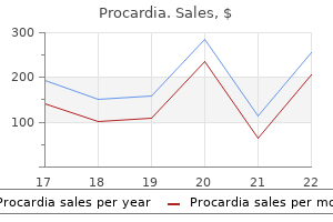
Procardia 30 mg order onlineEach linear hyaluronan molecule is associated with a lot of aggrecan molecules (more than 300) arteries to the head order procardia 30 mg fast delivery, which are certain to the hyaluronan by link proteins at the N terminus of the molecule to type massive proteoglycan aggregates 4 blood vessels of the heart procardia 30 mg order with amex. The entrapment of those aggregates within the intricate matrix of collagen fibrils is responsible for the unique biomechanical properties of hyaline cartilage. Multiadhesive glycoproteins, additionally referred to as noncollagenous and nonproteoglycan-linked glycoproteins, affect proteoglycan monomer (aggrecan) interactions between the chondrocytes and the matrix molecules. Multiadhesive glycoproteins have medical worth as markers of cartilage turnover and degeneration. Hyaline cartilage matrix is very hydrated to present resilience and diffusion of small metabolites. Much of this water is certain tightly to the aggrecan�hyaluronan aggregates, which create excessive osmotic swelling strain. These massive hydrodynamic domains within the matrix are accountable for imparting resilience to the cartilage. Some of the water is certain loosely sufficient to enable diffusion of small metabolites to and from the chondrocytes. In articular cartilage, each transient and regional modifications happen in water content during joint motion and when the joint is subjected to stress. A hyaluronan molecule forming a linear aggregate with many proteoglycan monomers is interwoven with a community of collagen fibrils. The proteoglycan monomer (such as aggrecan) consists of roughly 180 glycosaminoglycans joined to a core protein. Throughout life, cartilage undergoes continuous inner transforming as the cells substitute matrix molecules lost via degradation. Normal matrix turnover is decided by the power of the chondrocytes to detect changes in matrix composition. In addition, the matrix acts as a signal transducer for the embedded chondrocytes. Thus, stress hundreds applied to the cartilage, as in synovial joints, create mechanical, electrical, and chemical alerts that assist direct the synthetic exercise of the chondrocytes. As the body ages, nonetheless, the composition of the matrix modifications, and the chondrocytes lose their ability to reply to these stimuli. Chondrocytes are specialised cells that produce and maintain the extracellular matrix. This specimen was preserved in glutaraldehyde, embedded in plastic, and stained with H&E. The chondrocytes, particularly these within the higher a part of the photomicrograph, are nicely preserved. The cytoplasm is deeply stained, exhibiting a distinct and relatively homogeneous basophilia. This layer represents deposition of latest cartilage (appositional growth) on the floor of the prevailing hyaline cartilage. Mature chondrocytes with clearly seen nuclei (N) reside in the lacunae and are well preserved in this specimen. Growth from throughout the cartilage (interstitial growth) is mirrored by the chondrocyte pairs and clusters that are liable for the formation of isogenous teams (rectangles). When the chondrocytes are current in isogenous groups, they represent cells that have just lately divided. They additionally secrete metalloproteinases, enzymes that degrade cartilage matrix, allowing the cells to expand and reposition themselves within the growing isogenous group. Chondrocytes not only secrete the collagen present in the matrix but also the entire glycosaminoglycans and proteoglycans. In older, less energetic cells, the Golgi equipment is smaller; clear areas of cytoplasm, when evident, usually point out sites of extracted lipid droplets and glycogen shops. In such specimens, chondrocytes also display appreciable distortion resulting from shrinkage after the glycogen and lipid are misplaced during preparation of the tissue. Thus, the basophilia and metachromasia seen in stained sections of cartilage provide details about the distribution and relative focus of sulfated proteoglycans. It additionally has a decrease focus of sulfated proteoglycans and stains much less intensely than the capsular matrix. The interterritorial matrix is a area that surrounds the territorial matrix and occupies the space between teams of chondrocytes. Initially, most lengthy bones are represented by cartilage fashions that resemble the shape of the mature bone (Plate eight, web page 208). During the developmental course of, by which most of the cartilage is changed by bone, residual cartilage at the proximal and distal finish of the bone serves as growth sites known as epiphyseal growth plates (epiphyseal discs). The hyaline cartilage of developing tarsal bones might be changed by bone as endochondral ossification proceeds. In this early stage of development, synovial joints are being fashioned between creating tarsal bones. Note that nonarticulating surfaces of the hyaline cartilage fashions of tarsal bones are lined by perichondrium, which also contributes to the event of joint capsules. Also, a developing tendon (T) is obvious in the indentation of the cartilage seen on the left side of the micrograph. A disc of hyaline cartilage-the epiphyseal plate- separates the more proximally situated epiphysis from the funnel-shaped diaphysis situated distal to the plate. The articular cartilage on the surface of the epiphysis contributes to the synovial joint and can additionally be composed of hyaline cartilage. The cartilage of the epiphyseal plate disappears when lengthwise growth of the bone is accomplished, but the articular cartilage stays throughout life. Hyaline cartilage additionally exists in the grownup because the skeletal unit in the trachea, bronchi, larynx, and nostril. A firmly attached connective tissue, the perichondrium, surrounds hyaline cartilage. Hyaline cartilage that covers the articular surfaces of movable joints is termed articular cartilage. In common, the the perichondrium is a dense irregular connective tissue composed of cells which might be indistinguishable from fibroblasts. In many respects, the perichondrium resembles the capsule that surrounds glands and many organs. When actively rising, the perichondrium seems divided into an inside mobile layer, which gives rise to new cartilage cells, and an outer fibrous layer. Articular cartilage is a remnant of the original hyaline cartilage template of the developing bone, and it persists all through grownup life. This diagram shows the organization of the collagen community and chondrocytes within the varied zones of articular cartilage. Also, this zone is lighter staining than the matrix of the more superficial zones. Collagen fibrils are much less organized and are arranged in a somewhat indirect orientation to the surface.
Diseases - Filariasis
- Porphyria, hereditary coproporphyria
- Spondylohypoplasia arthrogryposis popliteal pteryg
- Fanconi anemia type 1
- Short rib-polydactyly syndrome, Majewski type
- Malonyl-CoA decarboxylase deficiency
- Phosphoglycerate kinase deficiency
30 mg procardia otcAdipose tissue of the epicardium contains branches of the coronary arteries and tributaries of the coronary veins coronary heart 70s purchase procardia 30 mg on-line. The latter is steady with the the fibrosa types the core of the valve and incorporates fibrous extensions from the dense irregular connective tissue of the skeletal rings of the center cardiovascular center of south florida cheap procardia 30 mg. The spongiosa is free connective tissue situated on the atrial or blood vessel side of every valve. It consists of loosely arranged collagen and elastic fibers infiltrated with massive numbers of proteoglycans. The spongiosa acts as a shock absorber to dampen vibrations related to the closing of the valve. In the aortic and pulmonary valves, spongiosa located on the blood vessel aspect is called arterialis. This photomicrograph shows a sagittal section of the posterior wall of the left atrium and left ventricle. The ventricular wall consists of three layers: (1) endocardium (arrowheads), (2) myocardium, and (3) epicardium. The seen blood vessels lie in the epicardium and are surrounded by adipose tissue. This excessive magnification of the world indicated by the rectangle reveals the attribute options of the internal floor of the center. The ventricularis is instantly adjacent to the ventricular or atrial floor of each valve and is roofed with endothelium. Intrinsic Regulation of Heart Rate Contraction of the heart is synchronized by specialized cardiac conducting cells. The surfaces of the valve are exposed to blood, and the cusps are thin enough to enable nutrients and oxygen to diffuse from the blood. These situations, identified collectively as valvular heart disease, embody rheumatic coronary heart illness, vegetative endocarditis, degenerative calcific aortic valve stenosis, and mitral annular calcification. For instance, rheumatic fever causes inflammation of the heart valves (valvulitis). Inflammation induces angiogenesis in the valve and vascularization in the usually avascular layers of the valve. These adjustments mostly affect the mitral valve (65% to 70%) and aortic valve (20% to 25%). This inflammation can result in progressive alternative of elastic tissue by irregular plenty of collagen fibers, causing the valve to thicken. The valves turn into rigid and inflexible, which impacts their capability to open and shut. Cardiac muscle can contract in a rhythmic manner without any direct stimulus from the nervous system. The electrical exercise (impulses) that ends in the rhythmic pulsations of the center is initiated and propagated by the conducting system of the center. The fee of depolarization of cardiac muscle varies in several elements of the conducting system; the quickest is within the atria, the slowest within the ventricles. The contraction cycle of the center is initiated within the atria, forcing blood into the ventricles. A wave of contraction within the ventricles then begins on the apex of the heart, forcing blood from the heart into the aorta and pulmonary trunk. The conducting system of the center consists of two nodes-the sinoatrial (or sinu-atrial) node and the atrioventricular node-and a series of conduction fibers or bundles (tracts). This photograph reveals a sagittal part of the posterior wall of the left ventricle and the posterior cusp of the mitral valve. The chordae tendineae extend from the papillary muscle to the ventricular aspect of the mitral valve cusp. Glistening internal floor of the guts represents the endocardium; the outer surface of the myocardium is roofed by the epicardium. This photomicrograph reveals a bit by way of one of the two cusps of the mitral valve. Beginning on the atrial side (top of the image), the first layer underlying the endothelium is the spongiosa-not well developed in this a part of the cusp. The second layer is the fibrosa, which types the majority of the dense connective tissue within the core of the valve. The third layer, the ventricularis, is fashioned by dense connective tissue containing layers of elastic and collagen fibers. The components of the conducting system convey impulses at a fee roughly four times sooner than the cardiac muscle fibers and are the one parts that may convey impulses across the fibrous skeleton. In complete coronary heart block when the conduction of electric impulses to the ventricles is interrupted, the ventricles will beat at their very own rate of about 30 to forty beats per minute, driven by depolarization of Purkinje fibers. Purkinje fibers have the slowest rate of intrinsic depolarization of the whole conducting system. Electrodes report electrical exercise of the heart by measuring voltage differences between completely different points. The nuclei are spherical and are larger than the nuclei of the cardiac muscle cells in the myocardium. Because of the considerable size of the cells, the nuclei are sometimes not included in the section. Because of the saved glycogen, Purkinje fiber cells are extra immune to hypoxia than are ventricular muscle cells. This photomicrograph reveals a Mallory-Azan�stained part of the ventricular wall of a human coronary heart. The upper two-thirds of the micrograph is occupied by the endocardium (E) containing a thick layer of Purkinje fibers. The free luminal floor of the ventricle (top) is roofed by endothelium and an underlying layer of subendothelial connective tissue (stained blue). The Purkinje fibers comprise giant amounts of glycogen, which seem as homogeneous, pale-staining areas that occupy the middle portion of the cell surrounded by the myofibrils. The nuclei (N) are spherical and are bigger than the nuclei of the cardiac muscle cells within the myocardium (M). They are regularly surrounded by the lighter stained cytoplasm, which represents the juxtanuclear region of the cell. Because of the appreciable measurement of the Purkinje cells, the nuclei are sometimes not included in the section. Systemic Regulation of Heart Function As talked about above, the heart beats independently of any nervous stimulation. This spontaneous rhythm of the center could be altered by nerve impulses from both sympathetic and the parasympathetic nerve supply to the heart originates within the vagus nerve (cranial nerve X). Presynaptic parasympathetic fibers synapse with postsynaptic neurons inside the coronary heart. The release of the neurotransmitter acetylcholine from the terminals of these fibers slows the guts rate (an impact known as bradycardia), reduces the pressure of the heartbeat, and constricts the coronary arteries of the guts. The sympathetic presynaptic fibers that offer the guts originate in the lateral horns on the stage of the T1 to T6 segments of the spinal twine.
Buy cheap procardia 30 mg on-lineThe alveolar ducts terminate in alveolar sacs cardiovascular disease prevalence discount procardia 30 mg on-line, enlarged areas surrounded by clusters of alveoli that open into the spaces lightcycler capillaries 20 l from roche procardia 30 mg cheap without a prescription. This consists of the alveolar epithelial cells and their basal lamina, the basal lamina of the underlying capillary endothelium and the endothelial cells, themselves, and another connective tissue elements that will lie between the two basal laminae. Some basal cells are nonetheless current, thus the designation pseudostratified columnar. Elsewhere, the epithelium might be ciliated simple columnar, and just before it turns into a respiratory bronchiole, the epithelium could embrace cuboidal or low columnar nonciliated cells. Characteristically, the wall of the respiratory bronchiole consists of alternating thick and thin regions. The thick areas are much like the wall of the bronchiole except that cuboidal Clara cells instead of columnar epithelium type the surface. The thin regions have a wall similar to the alveolar wall; this is considered beneath. The respiratory bronchiole proven in lower left determine is barely extra distal than the realm seen in top proper determine. Structurally, it exhibits essentially the same features as these seen in upper right determine besides that there are fewer Clara cells and the smooth muscle is considerably thinner. The central element of the alveolar wall is the capillary (C) and, in certain areas, related connective tissue. On each side, the place it faces the alveolus (A), a flat squamous cell is interposed between the capillary and the air areas. In some locations, the kind I cell is separated from the capillary endothelial cell by a single basal lamina shared by the 2 cells. This is the skinny portion of the alveolar�capillary complex, readily seen in the upper part of the determine (arrows). Elsewhere, connective tissue is interposed between the pneumocyte kind I cell and the endothelial cell of the capillary; every of these epithelial cells retains its own basal lamina. This cell sometimes shows a rounded (rather than flattened) form, and the nucleus is surrounded by a noticeable amount of cytoplasm, a few of which may appear clear. The septal cell produces a surface-active agent totally different from that of the Clara cell, which also acts in allowing the lung to expand. The kidneys play an important role in body homeostasis by conserving fluids and electrolytes and by disposing metabolic waste. Like the lungs and liver, the kidneys retrieve essential materials and dispose of wastes. To maintain homeostasis, kidneys conserve water, electrolytes, and certain metabolites. The kidneys are important in maintaining constant plasma pH by regulating acid�base balance, which is achieved by excreting hydrogen ions when bodily fluids turn into too acidic or excreting bicarbonates when bodily fluids turn into too fundamental. The kidneys play an essential role in regulating and sustaining the composition and volume of extracellular fluid. The kidneys are highly vascular organs; they obtain roughly 25% of the cardiac output. In the human physique, vitamin D is derived from two sources: � Skin, in which vitamin D3 (cholecalciferol) is quickly produced by the motion of ultraviolet gentle on the precursor 7-dehydrocholesterol. Typically, half-hour to 2 hours of sunlight publicity per day can present enough vitamin D to fulfill daily body requirements for this vitamin. In the blood, vitamin D3 is certain to vitamin D�binding protein and transported to the liver. The associated compound vitamin D2 (ergocalciferol) undergoes the identical conversion steps as vitamin D3 and produces the identical biologic effects. Patients with end-stage persistent kidney illnesses have inadequate conversion of vitamin D into active metabolites leading to vitamin D3 deficiency. In adults, vitamin D3 deficiency is manifested by impaired bone mineralization and reduced bone density. Therefore, patients with persistent kidney illnesses, particularly those on extended renal hemodialysis are sometimes supplemented with vitamin D3 and calcium to avoid extreme disturbance of calcium homeostasis due to secondary hyperparathyroidism, a situation prevalent in these sufferers. Vitamin D3 deficiency in childhood leads to rickets, a disease that causes abnormal bone ossification. Initially, plasma is separated from the cells and enormous proteins to produce a glomerular ultrafiltrate of the blood or major urine, which is then modified by selective resorption and specific secretion by the cells of the kidney. The final urine accommodates water and electrolytes in addition to waste products, similar to urea, uric acid, and creatinine, and breakdown merchandise of varied substances. They prolong from the 12th thoracic to the third lumbar vertebrae, with the right kidney positioned barely lower. On the upper pole of each kidney, embedded inside the renal fascia and a thick protective layer of perirenal adipose tissue, lies an adrenal gland. The medial border of the kidney is concave and contains a deep vertical fissure, called the hilum, through which the renal vessels and nerves move and the expanded, funnel-shaped origin of the ureter, referred to as the renal pelvis, exits. Although not shown within the illustration, the house between and round these structures is filled largely with unfastened connective tissue and adipose tissue. Synthesis and secretion of the acid protease renin, an enzyme concerned in charge of blood pressure and blood volume. Renin is produced by juxtaglomerular cells and cleaves circulating angiotensinogen to launch angiotensin I (see pages 713�714). Cortex and Medulla Examination with the naked eye of the reduce face of a fresh, hemisected kidney reveals that its substance may be divided into two distinct regions: hilum renal vein medullary rays � � Cortex, the outer reddish-brown part Medulla, the much lighter colored inside half minor calyx renal artery renal pelvis medulla the colour seen within the reduce surface of the unfixed kidney displays the distribution of blood in the organ. Approximately 90% to 95% of the blood passing through the kidney is within the cortex; 5% to 10% is within the medulla. The nephron is the basic useful unit of the kidney and is described in a following part. They constitute the start segment of the nephron and comprise a unique capillary community called a glomerulus. Their name displays their look, as the striations appear to radiate from the medulla. The diagram represents a hemisection of a kidney, revealing its structural group. The areas between medullary rays comprise the renal corpuscles, the convoluted tubules of the nephrons, and the connecting tubules. Each nephron and its connecting tubule (which connects to a accumulating duct in the medullary ray) type the uriniferous tubule. The medulla is characterised by straight tubules, amassing ducts, and a particular capillary community, the vasa recta. This photomicrograph of a Mallory-Azan�stained part reveals the capsule (cap) and a half of the underlying cortex. The fibroblasts in this a half of the capsule are comparatively few in number; their nuclei seem as slim, elongate, red-staining profiles in opposition to a blue background representing the stained collagen fibers.
30 mg procardia cheap with amexOne type of spindle cell cardiovascular fitness articles 30 mg procardia cheap overnight delivery, the nuclear bag fiber cardiovascular disease in children 30 mg procardia order visa, incorporates an aggregation of nuclei in an expanded midregion; the other type, known as a nuclear chain fiber, has many nuclei organized in a series. A typical muscle spindle consists of two to 4 nuclear bag fibers and roughly six to eight nuclear chain fibers. The muscle spindle transmits details about the degree of stretching in a muscle. When skeletal muscle is stretched, nerve endings of sensory nerves become activated and convey sensory information about muscle length and velocity of stretch. In addition, spindle cells obtain motor (efferent) innervation from the spinal wire and mind through two forms of motor efferent (type) nerve fibers, which are thought to regulate the sensitivity of the stretch receptors. Muscle spindles convey their impulses to the central nervous system, which in turn modulates the activity of motor neurons innervating that specific muscle. Such useful models precisely regulate contractions of parts of the muscle by creating "fixation factors" within the muscle tissue. Similar encapsulated receptors, Golgi tendon organs, are discovered in the tendons of muscle and reply to elevated tension on the muscle. These receptors contain solely sensory (afferent, Ib) nerve fibers, and they monitor muscle rigidity (or the force of contraction) within an optimum vary. Development, Repair, Healing, and Renewal Development of myogenic stem cell lineage is decided by expression of various myogenic regulatory components. Sensory Innervation Encapsulated sensory receptors in muscular tissues and tendons are examples of proprioreceptors. These receptors are part of the somatic sensory system that gives information about the diploma of stretching and rigidity in a muscle. The muscle spindle is a specialised stretch receptor positioned within the skeletal muscle. The muscle spindle is a specialized stretch receptor found in all skeletal muscular tissues; it consists of two forms of modified muscle Myoblasts are derived from a self-renewing inhabitants of multipotential myogenic stem cells that originate in the embryo from unsegmented paraxial mesoderm (cranial muscle progenitors) or segmented mesoderm of somites (epaxial and hypaxial muscle progenitors). It is thought that MyoD preferentially upregulates myostatin gene expression and controls myogenesis during not only the embryonic and fetal durations but also postnatal levels of improvement. Each spindle contains approximately two to four nuclear bag fibers and 6 to eight nuclear chain fibers. In the nuclear bag fibers, the muscle fiber nuclei are clumped in the expanded central portion of the fiber, hence the name bag. In distinction, the nuclei concentrated within the central portion of the nuclear chain fibers are organized in a series. The afferent nerve fibers respond to excessive stretching of the muscle, which in flip inhibits the somatic motor stimulation of the muscle. The efferent nerve fibers regulate the sensitivity of the afferent endings in the muscle spindle. Photomicrograph of a cross-section of a muscle spindle, exhibiting two bundles of spindle cells within the encapsulated, fluid-filled receptor. In one bundle, a quantity of of the spindle cells are minimize on the stage that reveals their nuclei. The exterior capsule of the muscle spindle and the adjoining perimysium can be seen as a faint double-layer boundary of the receptor. Immediately above and outdoors of the muscle spindle is a nerve that could be supplying the spindle. The flocculent material within the capsule consists of precipitated proteoglycans and glycoproteins from the fluid that stuffed the spindle before fixation. The hypermuscular phenotypes noticed on inactivation of the myostatin gene in animals and humans have confirmed the position of myostatin as a adverse regulator of skeletal-muscle improvement. Pharmacologic manipulation of myostatin expression might additionally result in the development of latest therapeutic approaches in a wide range of musculoskeletal pathologies. Secondary myotubes proceed to be shaped by sequential fusion of myoblasts into the already-formed secondary myotubes at random positions alongside their length. In the mature multinucleated muscle fiber, the nuclei are all within the peripheral sarcoplasm, simply inside the plasma membrane. Some nuclei that seem to belong to the skeletal muscle fiber are nuclei of satellite cells. Developing muscle accommodates two forms of myoblasts: � Early myoblasts are answerable for the formation of major myotubes, chain-like structures that reach between tendons of the developing muscle. Primary myotubes noticed within the mild microscope exhibit a chain of multiple central nuclei surrounded by myofilaments. Late in fetal development, the multipotential myogenic stem cell inhabitants generates satellite tv for pc cells, which are characterized by the expression of paired box transcription factor member of the family Pax7. Therefore, in a developing muscle, a pool of undifferentiated cells which have the potential to undergo myogenic differentiation is preserved. Satellite cells are small with scant cytoplasm, they usually make up 2% to 7% of all nuclei related to a single muscle fiber. This photomicrograph reveals a cross-section (on the left) and a longitudinal section (on the right) of creating skeletal muscle fibers within the stage of secondary myotubes. These myotubes are formed by sequential fusion of myoblasts, forming elongated tubular structures. Note that the myotubes have a small diameter and extensively spaced, centrally positioned nuclei that steadily turn into displaced into the cell periphery by the increased variety of newly synthesized myofilaments. In the mature multinucleated muscle fiber (upper left), all nuclei are positioned in the peripheral sarcoplasm, simply inside the plasma cell membrane. Myogenic precursor cells then downregulate Pax7 and differentiate, giving rise to new myoblasts. As lengthy because the external lamina stays intact, the myoblasts fuse within the exterior lamina to kind myotubes, which then mature into a brand new fiber. In contrast, if the exterior lamina is disrupted, fibroblasts repair the injured web site, with subsequent scar tissue formation. Muscular dystrophies are characterized by progressive degeneration of skeletal muscle fibers, which places a continuing demand on the satellite tv for pc cells to replace the degenerated fibers. New experimental data indicate that, throughout this course of, extra myogenic cells are recruited from the bone marrow and supplement the out there satellite cells. The fee of degeneration exceeds the rate of regeneration, nevertheless, leading to loss of muscle operate. A future therapy technique for muscular dystrophies might include the transplantation of satellite tv for pc cells or their myogenic bone marrow counterparts into damaged muscle. Each satellite cell has a single nucleus with a chromatin community denser and coarser than that of muscle cell nuclei. However, after muscle tissue harm, some satellite tv for pc cells are activated and turn out to be myogenic contractile filaments as skeletal muscle. Therefore, cardiac muscle cells and the fibers they kind exhibit cross-striations evident in routine histologic sections.
Asian Water Plantain (Water Plantain). Procardia. - What is Water Plantain?
- Dosing considerations for Water Plantain.
- Are there safety concerns?
- Bladder and urinary tract diseases.
- How does Water Plantain work?
Source: http://www.rxlist.com/script/main/art.asp?articlekey=96365
Buy generic procardia 30 mgUnlike the cells produced by mitosis 10 cardiovascular risk factors procardia 30 mg with amex, that are genetically equivalent to the mother or father cell heart disease symptoms women procardia 30 mg discount free shipping, the cells produced by meiosis are genetically unique. The chromosomes bear movement to finally align their centromeres along the equator of the spindle. The sister chromatids, held collectively by cohesin complexes and by the centromere, remain together. A maternal or paternal member of each homologous pair, now containing exchanged segments, strikes to every pole. Under normal physiologic circumstances (homeostasis), the charges of cell division and cell dying are related. If the rate of cell demise is larger than that of cell division, then a web lack of cell quantity will occur. When the scenario is reversed and the speed of cell division is greater than the speed of cell dying, then the web achieve in cell number shall be outstanding, resulting in a variety of problems of cell accumulation. Cell demise might occur because of acute cell damage or an internally encoded suicide program. Cell dying may outcome from accidental cell harm or mechanisms that cause cells to self-destruct. It happens when cells are exposed to an unfavorable bodily or chemical setting. Under physiologic situations, harm to the plasma membrane may also be initiated by viruses, or proteins known as perforins. Today, the term programmed cell demise is applied extra broadly to any sort of cell death mediated by an intracellular demise program, irrespective of the trigger mechanism. During apoptosis, cells that are no longer needed are eradicated from the organism. This course of could occur during normal embryologic growth or other normal physiologic processes, similar to follicular atresia within the ovaries. Cells can initiate their own death via activation of an internally encoded suicide program. Apoptosis is characterized by controlled autodigestion, which maintains cell membrane integrity; thus, the cell "dies with dignity" with out spilling its contents and damaging its neighbors. As a result of cell injury, harm to the cell membrane leads to an inflow of water and extracellular ions. As a result of the ultimate word breakdown of the plasma membrane, the cytoplasmic contents, including lysosomal enzymes, are released into the extracellular house. In apoptosis, the cell is an lively participant in its personal demise ("mobile suicide"). For example, cell demise mediated by cytotoxic T lymphocytes combines some features of both necrosis and apoptosis. Nuclear chromatin then aggregates, and the nucleus may divide into a number of discrete fragments bounded by the nuclear envelope. The cytoskeletal components turn out to be reorganized in bundles parallel to the cell floor. Loss of mitochondrial function is attributable to modifications within the permeability of the mitochondrial membrane channels. The integrity of the mitochondrion is breached, the mitochondrial transmembrane potential drops, and the electron-transport chain is disrupted. Thus, many researchers view mitochondria either as the "headquarters for the leader of a crack suicide squad" or as a "high-security jail for the leaders of a army coup. These membrane-bounded vesicles originate from the cytoplasmic bleb containing organelles and nuclear material. The removing of apoptotic our bodies is so environment friendly that no inflammatory response is elicited. In necrosis (left side), breakdown of the cell membrane results in an influx of water and extracellular ions, inflicting the organelles to endure irreversible changes. Lysosomal enzymes are released into the extracellular house, inflicting injury to neighboring tissue and an intense inflammatory response. Apoptotic bodies are later eliminated by phagocytotic cells with out inflammatory reactions. Apoptosis may also be inhibited by signals from other cells and the encircling setting through so-called survival components. These include progress elements, hormones similar to estrogen and androgens, impartial amino acids, zinc, and interactions with extracellular matrix proteins. However, an important regulatory function in apoptosis is ascribed to inner signals from the Bcl-2 (B-cell lymphoma 2) family of proteins. Members of this household consist of antiapoptotic and proapoptotic members that decide the life or demise of a cell. The proapoptotic members of the Bcl-2 family of proteins include Bad (Bcl-2�associated death promoter), Bax (Bcl-2�associated X protein), Bid (Bcl-2�interacting domain) and Bim (Bcl-2�interacting mediator of cell death). These proteins work together with each other to suppress or propagate their own activity by performing on the downstream activation of varied executional steps of apoptosis. Note the areas containing condensed heterochromatin adjacent to the nuclear envelope. The heterochromatin in one of the nuclear fragments (left) begins to bud outward by way of the envelope, initiating a model new round of nuclear fragmentation. Note the reorganization of the cytoplasm and budding of the cytoplasm to produce apoptotic bodies. These bodies will ultimately be phagocytosed by cells from the mononuclear phagocytotic system. Signals from an intercellular matrix are sensed by integrins that kind an integral part of anchoring cell-to-extracellular matrix junctions (see web page 142). Defects in these signaling pathways result in anoikis, which is triggered by the activation of the proapoptotic Bcl-2 household of proteins. This resistance is as a result of of varied mechanisms that embody changes within the integrin receptor types, activation of antiapoptotic factors, oncogene activation, and growth issue receptor signaling. Other Forms of Programmed Cell Death Several forms of programmed cell death had been lately identified that differ from apoptosis or necrosis. They embody the following: � Autophagy is a regulated mobile process that allows cells to turn over their contents by lysosomal degradation of their very own parts. This vacuole, known as an autophagosome, initially devoid of any lysosomal enzymes, fuses with lysosomes and initiates digestion. For an in depth description of three pathways utilized in autophagy, see pages 41 to forty three. Both exterior and inside stimuli can trigger apoptosis by activating the enzymatic caspase cascade. Failure to arrest the cell cycle before mitosis occurs causes problems with chromosome separation, which triggers the apoptotic pathway and cell dying. Paraptosis is an alternate, nonapoptotic cell demise that might be induced by development issue receptors. On a cellular degree, paraptosis is characterized by the formation of multiple giant vacuoles inside the cell cytoplasm along with mitochondrial swellings.
Procardia 30 mg discount with visaIntermediate Filaments As famous cardiovascular medicine davenport purchase procardia 30 mg overnight delivery, the molecular structure of intermediate filaments is tissue-specific and consists of many different sorts of proteins heart disease widowmaker cheap procardia 30 mg amex. Several illnesses are brought on by defects in the proper meeting of intermediate filaments. Accumulation of keratin intermediate filaments forming intercellular inclusions is frequently associated with particular cell injuries. In alcoholic liver cirrhosis, hepatocytes exhibit such inclusions (arrows), that are known as Mallory our bodies. Lymphocytes and macrophages responsible for an intense inflammatory response encompass cells containing Mallory our bodies. Microtubules develop upward from the basal physique, pushing the cell membrane outward, and elongate to type the mature cilium. During mitosis, the place of centrioles determines the placement of mitotic spindle poles. For occasion, astral microtubules are shaped round each individual centriole in a star-like trend. This schematic drawing reveals the orientation of the mitotic spindle in a traditional cell undergoing mitosis. Note the positions of the centrioles and the distribution of the spindle microtubules. In a cell that lacks centrioles, mitosis happens and a mitotic spindle containing only kinetochore microtubules is fashioned. However, each poles of the mitotic spindle lack astral microtubules, which place the spindle in the correct plane throughout mitosis. In the centriolar pathway, a pair of current centriole serves as an organizing middle for the duplication of latest centrioles. Utilizing this pathway, ciliated cells have the power to assemble giant variety of centrioles in the vicinity of an old mature centriole. In the acentriolar pathway, which performs a significant position in the formation of basal bodies in ciliated cells, new centrioles are formed de novo from fibrous granules located in shut proximity of nonmicrotubular constructions called deuterosomes. Both pathways give rise to procentrioles, which mature as they migrate to the appropriate website near the apical cell membrane, the place they turn out to be basal our bodies. The three microtubules of the triplet are fused, with adjoining microtubules sharing a typical wall. The innermost or A microtubule is an entire ring of 13 protofilaments containing - and -tubulin dimers; the middle and outer B and C microtubules, respectively, appear C-shaped because they share tubulin dimers with each other and with the A microtubule. The proximal a part of the lumen (close to the nucleus) is lined by -tubulin, which supplies the template for the arrangement of the triplet microtubules. In addition, a household of newly found -, -, -, and -tubulin molecules in addition to pericentrin protein complexes have also been localized with the centrioles. Other proteins, such as protein p210, type a ring of molecules that appears to link the distal end of the centriole to the plasma membrane. Centrosome duplication is synchronized with the cell-cycle events and linked to the method of ciliogenesis. Since each daughter cell receives just one pair of centrioles after cell division, the daughter cells should duplicate existing centrioles previous to cell division. In most somatic cells, duplication of centrioles begins near the transition between the G1 and S phases of the cell cycle. Note that the transverse-sectioned centriole in every of the pairs reveals the triplet configuration of microtubules. The decrease proper centriole represents a mid-longitudinal part, whereas the higher left centriole has also been longitudinally sectioned however along the airplane of its wall. In dividing cells, these connections participate in segregating the centrioles to each daughter cell. Their perform is to hyperlink the centriole to the mitotic spindle poles throughout mitosis. In human cells, the centrosome�nucleus connection seems to be maintained by filamentous buildings of cytoskeleton. A distinctive characteristic of mammalian centrioles is the distinction between particular person centrioles within the pair. In nondividing cells, centrioles are organized in pairs during which one centriole is aligned at a proper angle to the opposite. One centriole can be more mature (generated no much less than two cell cycles earlier) than the opposite centriole, which was generated within the earlier cell cycle. The mature centriole is characterised by the presence of satellites and appendages. The basic parts of each centriole are microtubule triplets that kind the cylindrical construction surrounding an inside lumen. The proximal part of the lumen is lined by -tubulin, which offers the template for nucleation and arrangement of the microtubule triplets. In some species, two protein bridges, the proximal and distal connecting fibers, join every centriole in a pair. The major cilium formation first happens throughout G1 phase during which the centrosome migrates towards the cell membrane and initiates the process of ciliogenesis. Necessary structural and transport proteins are acquired and activated to build major cilium axoneme (9 0) immediately on the top of the mature centriole. Duplication of centrioles begins near the transition between the G1 and S phases of the cell cycle, and the 2 centrioles are seen in S section. During the late G2 section, centrioles attain their full maturity, whereas the primary cilium is disassembled. This allows centrioles to migrate away from the cell membrane and participate within the mitotic spindle formation. Once cell division is full, the centrioles can proceed to ciliary reassembly in G1 phase. In most cells, duplication begins with the splitting of a centriole pair, adopted by the looks of a small mass of fibrillar and granular material on the proximal lateral end of every original centriole. Microtubules begin to develop within the mass of fibrous granules as it grows (usually during the S to late G2 phases of the cell cycle), showing first as a ring of nine single tubules, then as doublets, and eventually as triplets. As procentrioles mature in the course of the S and G2 phases of the cell cycle, every parent�daughter pair migrates across the nucleus. Before the onset of mitosis, centrioles with surrounding amorphous pericentriolar material position themselves on reverse sides of the nucleus and produce astral microtubules. In doing so, they define the poles between which the bipolar mitotic spindle develops. The important difference between duplication of centrioles throughout mitosis and during ciliogenesis is the reality that during mitosis, just one daughter centriole buds from the lateral facet of mother or father organelle, whereas during ciliogenesis, as many as 10 centrioles could develop across the parent centriole. Basal Bodies Development of cilia on the cell floor requires the presence of basal bodies, buildings derived from centrioles. The era of centrioles, which happens during the strategy of ciliogenesis, is responsible for the production of basal our bodies. The newly fashioned centrioles migrate to the apical surface of the cell and serve as organizing centers for the meeting of the microtubules of the cilium.
Procardia 30 mg buy without a prescriptionThese markers are the structural subunits TnI and TnT of the cardiac troponin complex capillaries growing 30 mg procardia discount fast delivery. Note that the nuclei in the longitudinally sectioned muscle cells seem elongate and also exhibit tapering ends cardiovascular disease environmental factors procardia 30 mg generic without prescription, thus matching the shape of the cell. In contrast, the nuclei in the cross-sectioned muscle cells are circular in profile. Also, a few of the cross-sectioned cells appear to lack a nucleus, a reflection that the part passed by way of one of many ends of the cell. In the previous, it was thought that after cardiac muscle cells are destroyed, they could not be replaced by new muscle cells. Recent research of hearts removed from individuals who had received transplants reveal nuclei undergoing mitosis. Perhaps in the future, a method might be developed that could induce human cardiac muscle to regenerate into wholesome tissue. The easy muscle cells, additionally called fibers, lack the striated sample found in skeletal and cardiac muscle. They range in size from 20 m within the partitions of small blood vessels to about 200 m in the wall of the intestine; they might be as massive as 500 m within the wall of the uterus throughout pregnancy. Small molecules or ions can pass from cell to cell by way of these junctions and provide communication hyperlinks that regulate contraction of the complete bundle or sheet of easy muscle. Smooth muscle cytoplasm stains somewhat evenly with eosin in routine H&E preparations because of the concentrations of actin and myosin that these cells include. The nuclei of clean muscle cells are positioned within the middle of the cell and sometimes have a corkscrew appearance in longitudinal section. This attribute is a result of contraction of the cell during fixation and is usually helpful in distinguishing clean muscle cells from fibroblasts in routine histologic sections. In the noncontracted cell, the nucleus appears as an elongated structure with tapering ends, lying in the center axis of the cell. When the nucleus is included in a cross-section of a smooth muscle fiber, it seems as a round or circular profile whether the cell is contracted or relaxed. Structure of Smooth Muscle Smooth muscle cells possess a contractile equipment of skinny and thick filaments and a cytoskeleton of desmin and vimentin intermediate filaments. The remaining sarcoplasm is full of thin filaments that type part of the contractile apparatus. Thick myosin filaments are scattered all through the sarcoplasm of a clean muscle cell. However, the structure of thick filaments in clean muscle is completely different than in skeletal muscle. This side-polar myosin filament additionally has no central "bare zone" however instead has asymmetrically tapered bare ends. This organization maximizes the interaction between thick and thin filaments, permitting the overlapped skinny filaments to be pulled over the whole size of the thick filaments. Several more proteins are associated with the contractile equipment and are important to initiation or regulation of the graceful muscle contractions. The bulk of the cytoplasm is occupied by thin (actin) filaments, which are simply recognizable at this magnification. The -actinin�containing cytoplasmic densities, or dense our bodies, are visible among the many myofilaments (arrows). It initiates the contraction cycle after its activation by Ca2 �calmodulin complicated. Calmodulin, a 17 kDa Ca2 -binding protein, is related to the TnC present in skeletal muscle, which regulates the intracellular concentration of Ca2. It may also, with caldesmon, regulate its phosphorylation and release from F-actin. These structures are distributed all through the sarcoplasm in a community of intermediate filaments containing the protein desmin. Note that vascular easy muscle incorporates vimentin filaments along with desmin filaments. The components of the contractile apparatus in easy muscle cells are the following. Dense our bodies present an attachment web site for thin filaments and intermediate filaments. Research means that the tropomyosin place on the actin filament is regulated by phosphorylation of myosin heads. Caldesmon (120 to 150 kDa) and calponin (34 kDa) are actin-binding proteins that block the myosin-binding web site. The motion of these proteins is Ca2 -dependent and is also managed by the phosphorylation of myosin heads. Dense bodies include quite lots of attachment plaque proteins, together with -actinin, which anchors each skinny filaments and intermediate filaments either instantly or indirectly to the sarcolemma. In support of this idea is the finding that dense bodies, although incessantly showing as small, isolated, irregular, electron-dense bodies, may seem as irregular linear constructions. Contraction in easy muscle tissue is initiated by a wide range of impulses, including mechanical, electrical, and chemical stimuli. The mechanisms that trigger contraction of clean muscle cells are very totally different from these of striated muscle. Smooth muscle has numerous sign transduction pathways that provoke and modulate smooth muscle contraction. The rectangle within the inset reveals portions of three easy muscle cells that seem at larger magnification in the massive electron micrograph. The -actinin�containing cytoplasmic densities (single arrows) usually seem as irregular lots, some of which are in touch with, and connected to , the plasma membrane. The cell within the center of the micrograph has been minimize in a aircraft closer to the cell floor and divulges these similar densities as a branching construction (double arrows). A three-dimensional mannequin of the cytoplasmic densities would reveal an anastomosing community. Higher magnification of cytoplasmic densities hooked up to the plasma membrane from the realm indicated by the rectangle. In addition, the pinocytotic vesicles could be observed in different phases of their formation. Electrical depolarizations can occur, corresponding to these throughout neural stimulation of smooth muscle. The release � of the neurotransmitters acetylcholine and norepinephrine from their synaptic nerve endings stimulates receptors positioned in the neuronal plasma membrane and changes the membrane potential. They have a helical parallel�antiparallel association of myosin molecules with their globular heads projecting from each ends of the filament.
Cheap 30 mg procardia with visaThis fusion produces a hybridoma coronary artery game procardia 30 mg buy with amex, an immortalized individual antibody-secreting cell line arteries arterioles capillaries venules veins 30 mg procardia with visa. To acquire monoclonal antibodies towards rat actin molecules, for instance, the B lymphocytes from the lymphatic organs of immunized rabbits have to be fused with myeloma cells. Both direct and oblique immunocytochemical methods are used to locate a target antigen in cells and tissues. The oldest immunocytochemistry method used for figuring out the distribution of an antigen inside cells and tissues is identified as direct immunofluorescence. Monoclonal antibodies conjugated with radioactive compounds are used to detect and diagnose tumor metastasis in pathology, differentiate subtypes of tumors and stages of their differentiation, and in infectious disease analysis to determine microorganisms in blood and tissue fluids. In current clinical studies, monoclonal antibodies conjugated with immunotoxins, chemotherapy agents, or radioisotopes have been used to deliver therapeutic agents to particular tumor cells within the body. Direct immunofluorescence strategies are actually being replaced by the oblique method due to suboptimal sensitivity. Indirect immunofluorescence provides much greater sensitivity than direct strategies and is usually referred to because the "sandwich" or "double-layer approach. Therefore, when the fluorescein is conjugated instantly with the precise main antibody, the method is direct; when fluorescein is conjugated with a secondary antibody, the tactic is oblique. The oblique method considerably enhances the fluorescence sign emission from the tissue. It can also be possible to conjugate polyclonal or monoclonal antibodies with other substances, similar to enzymes. In another variation, colloidal gold or ferritin (an iron-containing molecule) could be hooked up to the antibody molecule. These electron-dense markers may be visualized instantly with the electron microscope. In direct immunofluorescence, a fluorochrome-labeled primary antibody reacts with a particular antigen throughout the tissue pattern. Labeled buildings are then noticed within the fluorescence microscope in which an excitation wavelength (usually ultraviolet light) triggers the emission of one other wavelength. The size of this wavelength depends on the nature of the fluorochrome used for antibody labeling. Second, the secondary antibodies, that are fluorochrome labeled, react with the first antibodies. The visualization of labeled buildings within the tissue is similar in both methods and requires the fluorescence microscope. The conduct of microtubules (elements of the cell cytoskeleton) obtained from human breast tumor cells can be studied in vitro by measuring their nucleation activity, which is initiated by the centrosome. By use of oblique immunofluorescence techniques, microtubules have been labeled with a mixture of anti� -tubulin and anti� -tubulin monoclonal antibodies (primary antibodies) and visualized by secondary antibodies conjugated with fluorescein dye (fluorescein isothiocyanate�goat anti-mouse immunoglobulin G). The antigen�antibody response, carried out instantly on the glass coverslip, leads to visualization of tubulin molecules liable for the formation of greater than 120 microtubules seen on this picture. They originate from the centriole and extend outward approximately 20 to 25 m in a uniform radial array. The amplified transcripts obtained during these procedures are often detected using labeled complementary nucleotide probes in standard in situ hybridization techniques. For instance, a probe hybridized to metaphase chromosomes can be used to identify the chromosomal place of a gene. The frequency of chromosome translocations in lymphocytes is proportional to the absorbed radiation dose. Autoradiography Autoradiography makes use of a photographic emulsion positioned over a tissue section to localize radioactive materials within tissues. Hybrids are detected most frequently utilizing a radioactive label hooked up to one part of the hybrid. Binding of the probe and sequence can take place in an answer or on a nitrocellulose membrane. Digoxigenin and biotin are detected by immunocytochemical and cytochemical methods, respectively. The right nucleus is from a traditional amniotic fluid specimen and reveals two green and two orange indicators, which indicates two copies of chromosomes thirteen and 21, respectively. The nucleus on the left has three orange signals, which indicate trisomy 21 (Down syndrome). The radioactivity is then traced to localize the bigger molecules in cells and tissues. Labeled precursor molecules can be injected into animals or introduced into cell or organ cultures. Sections of specimens that have incorporated radioactive materials are mounted on slides. In the dark, the slide is often dipped in a melted photographic emulsion, thus producing a thin photographic film on the surface of the slide. After appropriate exposure in a light-tight field, normally for days to weeks, the exposed emulsion on the slide is developed by normal photographic methods and completely mounted with a coverslip. These grains could additionally be used simply to indicate the location of a substance, or they could be counted to provide semiquantitative information about the quantity of a given substance in a particular location. Some of the cells exhibit aggregates of metallic silver grains, which appear as small black particles (arrows). Over time, the lowenergy radioactive particles emitted from the [3H]thymidine strike silver halide crystals in a photographic emulsion overlaying the specimen (exposure) and create a latent image (much like gentle putting photographic movie in a camera). During photographic improvement of the slide with its overlaying emulsion, the latent image, really the activated silver halide within the emulsion, is decreased to the metallic silver, which then appears as black grains in the microscope. Electron microscopic autoradiograph of the apical region of an intestinal absorptive cell. Note that the silver grains are concentrated over apical invaginations (inv) and early endosomal tubular profiles (tub). A specimen to be examined with the bright-field microscope should be sufficiently thin for gentle to pass by way of it. The resolving energy of the human eye-that is, the space by which two objects must be separated to be seen as two objects (0. The function of a microscope is to amplify a picture to a level at which the retina can resolve the knowledge that might in any other case be beneath its limit of decision. Resolving energy is the ability of a microscope lens or optical system to produce separate images of closely positioned objects. Examination of a Histologic Slide Preparation within the Light Microscope Organs are three-dimensional, whereas histologic sections are only two-dimensional. This is the theoretical decision and, as talked about, is dependent upon all conditions being optimal. Various gentle microscopes are available for common and specialized use in modern biologic analysis. Their variations are based largely on such factors because the wavelength of specimen illumination, bodily alteration of the light coming to or leaving the specimen, and particular analytic processes that can be applied to the final picture. The microscope utilized by most college students and researchers is the bright-field microscope.
Order procardia 30 mg with amexAlso cardiovascular disease leading cause of death generic procardia 30 mg with visa, particular mesenchymal stem cells arrive to the location of harm from the encompassing gentle tissues and bone marrow capillaries in your body discount 30 mg procardia free shipping. Both fibroblasts and periosteal cells take part during this section of the therapeutic. Granulation tissue transforms into fibrocartilaginous soft callus, which supplies the fracture a steady, semirigid structure. Bony callus replaces fibrocartilage on the fracture site and permits for weight bearing. As the granulation tissue becomes denser, chondroblasts differentiate from the periosteal lining and the newly produced cartilage matrix invades the periphery of granulation tissue. While the callus is forming, osteoprogenitor cells of the periosteum divide and differentiate into osteoblasts. The newly fashioned osteoblasts start to deposit osteoid on the outer floor of the callus (intramembranous process) at a distance from the fracture. This new bone formation progresses towards the fracture site till new bone forms a bony sheath over the fibrocartilaginous callus. This low-magnification photomicrograph of a 3-week-old bone fracture, stained with H&E, exhibits parts of the bone separated from each other by the fibrocartilage of the delicate callus. In addition, the osteoblasts of the periosteum are involved in secretion of recent bony matrix on the outer floor of the callus. On the right of the microphotograph, the delicate callus is roofed by periosteum, which also serves as the attachment site for the skeletal muscle. Higher magnification of the callus from the area indicated by the upper rectangle in panel a reveals osteoblasts lining bone trabeculae. Most of the original fibrous and cartilaginous matrix at this site has been changed by bone. The early bone is deposited as an immature bone, which is later changed by mature compact bone. Higher magnification of the callus from the realm indicated by the decrease rectangle in panel a. A fragment of old bone pulled away from the fracture website by the periosteum is now adjoining to the cartilage. The cartilage will calcify and be replaced by new bone spicules as seen in panel b. In addition, endosteal proliferation and differentiation happen within the marrow cavity, and bone grows from both ends of the fracture towards the middle. When this bone unites, the bony union of the fractured bone, produced by the osteoblasts and derived from each the periosteum and endosteum, consists of spongy bone. As in normal endochondral bone formation, the spongy bone is gradually changed by woven bone. Bone reworking of the exhausting callus needs to happen so as to transform the newly deposited woven bone into a lamellar mature bone. It is typically accompanied by pain and swelling, and it leads to granulation tissue formation. The soft callus is shaped in approximately 2 to three weeks after fracture, and exhausting callus by which the fractured fragments are firmly united by new bone requires three to 4 months to develop. The process of bone transforming may last from a quantity of months to several years until the bone has utterly returned to its authentic form. Bone contributes to the skeleton, Bone which helps the physique, protects vital buildings, provides mechanical bases for physique movement, and harbors bone marrow. Long bones are tubular in form and include two ends (proximal and distal epiphyses) and a long shaft (diaphysis). Periosteum incorporates a layer of osteopro- osteoprogenitor cells and secrete osteoid, an unmineralized bone matrix that undergoes mineralization triggered by matrix vesicles. They talk with different osteocytes by a network of long cell processes occupying canaliculi, and they respond to mechanical forces utilized to the bone. Osteoclasts differentiate from hemopoietic progenitor cells; they resorb bone matrix during bone formation and transforming. Bone matrix incorporates mainly sort I collagen together with other noncollagenous proteins and regulatory proteins. Bone cavities are lined by endosteum, a single layer of cells that contains osteoprogenitor (endosteal) cells, osteoblasts, and osteoclasts. Bones articulate with neighboring bones by synovial joints, a movable con- nection. The articular surfaces that type contact areas between two bones are covered by hyaline (articular) cartilage. Compact bone lies exterior and beneath the periosteum, whereas an inner, sponge-like meshwork of trabeculae forms spongy bone. These concentric lamellar buildings are organized round an osteonal (Haversian) canal that incorporates the vascular and nerve supply of the osteon. The lacunae between concentric lamellae contain osteocytes that are interconnected with different osteocytes and the osteonal canal by way of canaliculi. Flat bones of the cranium, mandible, and clavicle develop by intramembranous ossification; all different bones develop by endochondral ossification. Next, osteoprogenitor cells surrounding this model differentiate into bone-forming cells that originally deposit bone on the cartilage surface (periosteal bony collar) and later penetrate the diaphysis to type the first ossification middle. Primary and secondary ossification centers are separated by the epiphyseal growth plate, offering a supply for model new cartilage concerned in bone development seen in children and adolescents. Epiphyseal progress plate has several zones (reserve cartilage, proliferation, hypertrophy, calcified cartilage, and resorption). Bone will increase in width (diameter) by appositional development of latest bone that occurs between the compact bone and the periosteum. Bone is constantly being transformed all through life by bone-remodeling models composed of osteoclasts and osteoblasts. Bone can restore itself after harm either by a direct (primary) or indirect (secondary) bone therapeutic course of. After damage, periosteal cells turn out to be activated to produce gentle (fibrocartilage) callus, which is subsequently changed by exhausting (bony) callus. Ca2 could additionally be removed from bone if the circulating stage of Ca2 within the blood falls under the critical worth. Bone serves as a storage web site for calcium and phosphate, which could be launched to the blood to preserve homeostatic ranges. Osteocytes reside in lacunae in the bone matrix and prolong fine mobile processes into canaliculi that join the lacunae, thus forming a steady community of cells throughout the mineralized tissue. Bones are organs of the skeletal system; bone tissue is the structural part of bones. Ground sections of bone are ready from bone that has not been fastened but merely allowed to dry. Thin slices of the dried bone are then reduce with a saw and additional ground to a thinness that permits viewing in a light-weight microscope. Slices may be treated with India ink to fill areas that have been formerly occupied by organic matter, for example, cells, blood vessels, and unmineralized matrix. A less complicated technique is to mount the ground specimen on a slide with a viscous medium that traps air in a few of the areas, as in the specimen on this plate.
|
