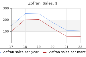
Proven 8 mg zofranIn a pilot study treatment 4s syndrome zofran 8 mg buy cheap on line, uterine artery ligation at the time of cesarean delivery appeared to improve shrinkage of fibroids postpartum [38] symptoms quiz buy cheap zofran 8 mg line, however further scientific research and risk/benefit analyses are wanted before such an intervention could be really helpful. Management of Patients with Prior Myomectomy Method of delivery and timing of cesarean supply. In the absence of strong proof of the absolute danger of rupture, it is recommended to take a conservative method and perform cesarean supply previous to the onset of labor if the myometrium was considerably compromised by earlier surgical procedure, corresponding to entry into the uterine cavity or near entry during a prior myomectomy or if numerous myomas had been eliminated. The magnitude of the risk of uterine rupture in pregnancies after myomectomy and particular criteria related to increased danger are difficult to ascertain due to the small number of circumstances reported and lack of element concerning the operative procedures performed. In a 2016 systematic evaluate of studies with a minimal of 5 circumstances of being pregnant after myomectomy, the general incidence of uterine rupture after myomectomy was 7/756 or 0. Six of the seven ruptures occurred in girls who had a prior laparoscopic myomectomy, which has been attributed to the technical challenge of laparoscopic suturing [41]. All ruptures occurred following myomectomy of an intramural fibroid, though this was not noted to be a major danger issue for uterine rupture. The uterine cavity was not entered throughout myomectomy in three instances; this info was not obtainable in the other 4 cases. The ruptures occurred at 24 (twins), 25, 30, 32, 36, 37, and 40 weeks of gestation; nevertheless, this finding may be biased by scheduled cesarean deliveries at time period. It is essential for the gynecologist who performs any type of myomectomy to clearly identify within the operative observe the quantity, measurement, and location of the tumor; the depth and number of uterine incisions; and any entrance of the uterine cavity. A recommendation to the lengthy run obstetrician of whether elective cesarean section or trial of labor for the attainable coming pregnancies is advisable. Prior hysteroscopic myomectomy of a submucosal fibroid may enhance the danger of irregular placentation, particularly placenta accreta because of the elevated probability of adhesion formation within the uterine cavity. Although the danger of placenta accreta after prior myomectomy appears to be low [25], data are sparse. Ultrasound screening for attainable placenta accreta in the late second or early third trimester is beneficial. Prevalence of uterine leiomyomas in the first trimester of pregnancy: An ultrasound-screening research. Leiomyomas at routine second-trimester ultrasound examination and adverse obstetric outcomes. Risk of uterine rupture and placenta accreta with prior uterine surgical procedure outside of the decrease segment. Epidural use of morphine in managing the pain of carneous degeneration of a uterine leiomyoma during being pregnant. In the United States, the annual financial burden of those tumors is evaluated to be $34. The rate of this disease could be affected by many components, together with race, physique mass index, family history, and ethnicities. Uterine fibroids are monoclonal tumors that arise from uterine smooth muscle (myometrium), and considered one of their characteristic features is their dependency on the ovarian steroids estrogen (E2) and progesterone (P4) [6]. Hormonal fluctuations influence fibroid development in the course of the being pregnant and postpartum periods. In addition to the hormonal response, these lesions are influenced by fifty five 56 Fibroids and Reproduction genetic aberrations [7,8]. The monoclonal origin of fibroids implies mutations of myometrial cells as the origin of the disease. It is properly studied that fibroids can be subdivided based on the presence of clonal chromosomal aberrations as, Estrogen has an important role in fibroid progress and growth, which explains the onset of signs at puberty and stops after menopause. Uterine fibroids have more estrogen and progesterone receptors than wholesome myometrial cells. Risk Factors of Uterine Fibroids Race the incidence of uterine fibroids is disproportionately higher in African American women compared with Caucasian girls. The prevalence of fibroids by age 50 years is greater than 80% in African American ladies, compared with 70% in Caucasian women. More studies on the etiology of uterine fibroids are required to establish the rationale for ethnic danger components in fibroids [14]. Reproductive Factors Parity and being pregnant play a protecting function in uterine fibroid growth, reducing the danger of uterine fibroids as a lot as 5 fold, first due to the decreased interval of exposure to unopposed estrogen and second due to the incidence of ischemia during parturition and uterine transforming. Obesity High body mass index is associated with a reasonable improve in danger of uterine fibroids. Obesity results in a rise within the conversion of adrenal androgens to estrogen, eventually leading to extra unbound energetic estrogen. Also, hyperinsulinemia that results in metabolic syndrome was found to be associated with an increased threat of uterine fibroids [16]. Vitamin D Deficiency Vitamin D deficiency was discovered to increase the incidence of uterine fibroids. Hormonal Effects Clinical and experimental research have proven that estrogen and progesterone stimulate the expansion of uterine fibroids. A prominent function of uterine fibroids is their dependency on the ovarian steroid hormones by way of the reproductive years. Progesterone is an endogenous steroid hormone associated with the menstrual cycle and being pregnant and has an important role in female replica and being pregnant. In uterine fibroids, progesterone controls many targets, which can play a vital role in uterine fibroid pathogenesis [22]. A direct practical hyperlink between progesterone and uterine fibroid improvement was proven in a mouse xenograft mannequin, reflecting characteristics of uterine fibroids by grafting human fibroid tissue beneath the renal capsule of immunodeficient mice. The mass of established fibroid xenografts reveals statistically vital diminishment in response to progesterone withdrawal, suggesting that the amount upkeep and development of fibroids are progesterone dependent [6]. Uterine Fibroid Management Treatment options of uterine fibroids embrace medical and surgical options. Women attempt to avoid the surgical option due to the dangers related to the operation and to save their uterus for future pregnancies. Medical choices are most well-liked; nevertheless, sometimes surgical intervention is considered the first alternative of treatment for uterine fibroids [24,25]. In this chapter, we cowl uterine fibroid remedy choices by addressing their impact on productivity maintenance. Hysteroscopic Myomectomy Hysteroscopy is a modality by which intrauterine pathologies may be identified and treated at the similar time. The extraction of submucosal fibroids is certainly one of the primary indications for hysteroscopic myomectomy. Surgical resection by hysteroscopy will increase pregnancy rates in submucosal fibroids, while myomectomy for intramural fibroids continues to be mentioned. Complexities include uterine perforation which may lead to close by organ damage, postoperative endometritis, and bleeding [24]. Laparoscopic myomectomy is secure and environment friendly, with a low complication fee of lower than 10%. Several complications have been seen in laparoscopic myomectomy, probably the most concerning and worrisome complication of laparoscopic myomectomy is a pregnancy-related uterine rupture.
Zofran 8 mg buy without prescriptionDiagnostic peritoneal lavage medications requiring aims testing zofran 8 mg generic on-line, though seldom used now premonitory symptoms zofran 8 mg purchase free shipping, is helpful when a patient is too unstable from a cardiopulmonary standpoint to tolerate imaging research. The finding of leukocytes within the lavage effluent in an unstable patient might, in excessive circumstances, constitute adequate grounds for laparotomy. Acute Appendicitis Acute appendicitis is a ubiquitous drawback, accounting for approximately 5% of all emergency division visits for patients beneath sixty five years of age21 and 30% of acute surgical abdominal emergencies in sufferers under 50 years of age worldwide. Acute cholecystitis is, generally, attributable to persistent obstruction of the cystic duct by a gallstone. Mild elevations in serum whole bilirubin and alkaline phosphatase ranges are common. Demonstration of gallstones could suggest biliary ache, whereas the finding of stones with gallbladder wall thickening, pericholecystic fluid, and pain on compression of the gallbladder with the ultrasound probe (sonographic Murphy sign) is essentially diagnostic of acute cholecystitis and has replaced hepatobiliary scintigraphy The Tokyo consensus criteria for the analysis of acute cholecystitis are proven in Table eleven. Patients with acute cholecystitis are greatest managed with cholecystectomy within 48 hours. Patients with scores greater than or equal to 5 ought to be evaluated by a surgeon or endure an imaging research to look for appendicitis. Typically, acute appendicitis begins with prodromal symptoms of anorexia, nausea, and imprecise periumbilical ache. Within 6 to eight hours, the pain migrates to the right decrease quadrant and peritoneal signs develop. In uncomplicated appendicitis, a low-grade fever to 38�C and mild leukocytosis are often present. Mesenteric adenitis may also be attributable to Yersinia enterocolitica (see Chapter 110). In pediatric patients, intussusception, congenital malformations similar to malrotation and Meckel diverticulum are the most typical causes. Imaging findings characteristic of cholecystitis Definite Diagnosis: One item in A and one item in B or C, when acute cholecystitis is suspected clinically Adapted from Hirota J, Takada T, Kawarada Y, et al. Diagnostic criteria and severity assessment of acute cholecystitis: Tokyo Guidelines. Nausea and vomiting happen quickly after the onset of pain and provide momentary reduction of discomfort. Fever, tachycardia, and signs of volume contraction, together with orthostatic hypotension, are frequent. Abdominal distention is frequent but could also be difficult to discern within the setting of obesity. Peritoneal indicators ought to be thought of an indication of intestinal ischemia or perforation. Plain films of the abdomen are diagnostic once they reveal dilated loops of small gut with air-fluid ranges and decompressed distal small bowel and colon. The appearance of the distinction within the cecum inside 12 hours predicts resolution of obstruction. They could complain of constipation or obstipation and are normally discovered to have a leukocytosis. In extreme cases, generalized peritonitis could also be current, making differentiation from different causes of a perforated viscus tough. Acute diverticulitis presents as a spectrum of disease from mild belly discomfort to gross fecal peritonitis, which is an acute surgical emergency. Patients with pancreatitis are usually more comfy sitting upright, leaning forward slightly. Abdominal examination reveals hypoactive bowel sounds and marked tenderness to percussion and palpation in the epigastrium. In uncommon patients, flank or periumbilical ecchymoses (Grey-Turner or Cullen sign, respectively) develop in the setting of pancreatic necrosis with hemorrhage. Elevated serum and urine amylase ranges are usually current throughout the first few hours of ache; serum lipase is also elevated. Depending on the cause and severity of pancreatitis, serum electrolyte, calcium, and blood glucose ranges and liver biochemical check and arterial blood fuel results may be abnormal. When organ failure persists for more than this time, or impacts multiple organ systems, severe acute pancreatitis is present. Patients on this last group usually require multidisciplinary care in an intensive care unit. A minority of sufferers with severe acute pancreatitis current with a profound intra-abdominal disaster, usually caused by thrombosis of the middle colic artery or proper colic artery, which journey in proximity to the head of the pancreas, with ensuing colonic infarction. Patients with a perforated peptic ulcer usually present with the sudden onset of extreme diffuse stomach ache. These patients may have the ability to specify the precise second of the onset of symptoms. In the usual case, the troubled affected person presents acutely with excruciating abdominal pain, usually with out prodromal symptoms. Abdominal examination reveals peritonitis, with rebound tenderness, guarding, and stomach muscular rigidity. In such instances, distinguishing perforated ulcer from other causes of a perforated viscus A perforated peptic ulcer ought to be suspected in any patient with the sudden onset of extreme stomach ache who presents with abdominal rigidity and free intraperitoneal air. Implementation of evidence-based follow modeled on the Surviving Sepsis Guidelines in Denmark decreased the 30-day. Acute Mesenteric Ischemia Acute mesenteric ischemia may end up from occlusion of a mesenteric vessel because of an embolus, which may emanate from an atheroma of the aorta or cardiac mural thrombus, or major thrombosis of a mesenteric vessel, normally at a website of atherosclerotic stenosis. Embolic occlusion had accounted for up to 50% of cases of mesenteric ischemia in the Eighties but, because of advances in the management of risk elements for embolization, accounts for no a couple of third of cases in the 2010s. It mostly affects the superior mesenteric, presumably because of the much less acute angle of the superior mesenteric artery origin from the abdominal aorta. Patients normally have a history of atherosclerotic disease, particularly in the coronary or cerebrovascular circulation. Nonocclusive mesenteric ischemia, also referred to as "low-flow" mesenteric ischemia, accounts for 10% of cases. The remaining 10% of cases of mesenteric ischemia end result from venous thrombosis, often associated with a thrombophilia, and focal segmental ischemia of the small gut (see Chapter 118). Because most circumstances of mesenteric ischemia occur in patients with vital cardiovascular comorbidities, outcomes are poor. On bodily examination, most patients seem acutely ill, but the presentation could additionally be subtle. Patients with acute embolic or thrombotic intestinal ischemia should be referred for instant revascularization and bowel resection. For sufferers with persistent signs, laparotomy for resection of infarcted intestine may be needed. The pain is tearing in nature and related to prostration, lightheadedness, and diaphoresis. If the patient survives transit to the hospital, shock is the commonest presentation.
Zofran 8 mg buy discount on lineEpidemiology Perioral dermatitis is most typical in younger women aged 15�25 years medicine side effects zofran 8 mg discount with amex, being fairly uncommon in males and in older ladies symptoms for pink eye zofran 8 mg effective. Pathogenesis the pathways leading to the development of perioral dermatitis are unclear. The basic historical past is of a preferred facial eruption which initially improves with corticosteroid use after which worsens or recurs upon continued use. Lesions typically involve the nasolabial grooves and, in severely affected sufferers, additionally affect the skin on the sides of the nose. Injury to tissue initiates a fancy mobile and biochemical exercise, which ends up in wound therapeutic. Non-healing wounds are a significant cause of morbidity and have an unlimited impact on healthcare expenditures. Factors affecting wound healing There are various local as nicely as systemic elements that have an result on the method of wound therapeutic. The presence of infection or of a foreign physique in the wound leads to impaired wound therapeutic. Advanced age, weight problems, stress, ailments corresponding to diabetes, continual renal illness, continual liver disease, smoking, medicines corresponding to glucocorticoids, and chemotherapeutic agents negatively influence wound healing. The prevalence of venous leg ulcers increases with increasing age and is extra frequent in males greater than 60 years of age and in girls. Pathogenesis the venous system in decrease extremity comprises superficial, communicating, and deep veins. It incorporates bicuspid valves, which ensure the unidirectional circulate of blood in path of the center. In a standing position, the stress within the venous system is equal to the hydrostatic strain in the legs (80 mm Hg). During muscle contraction, calf muscle tissue exert strain on deep veins, and blood is pushed upwards. Normal valve functioning maintains a unidirectional move of blood and prevents transmission of excessive venous pressure to the superficial venous system. Initially, the small blood vessels constrict after which platelets plug the endothelial gaps. White cells accumulate at the interface between the broken and the normal tissue. After about 18�24 hours, epidermal cells actively move on to the surface of the defect. Epidermal cells on the sides of the wound divide some hours later to make up for the loss. After 2�4 days, new capillaries start to sprout and vascularize the granulation tissue within the wound cavity. Damaged connective tissue is destroyed and removed by macrophages, and new collagen is secreted by fibroblasts. Myofibroblasts are fibroblastic cells that develop the power to contract and are liable for wound contraction. In the later phases, remodelling takes place so that the orientation of the dermal collagenous bundles to the unique strains of stress happens. The dermis finally develops a normal profile and the vasculature can be restored to regular contractility. Various theories have been proposed, one being the fibrin cuff principle, which postulates that increased intraluminal pressure in capillaries causes leakage of fibrinogen and formation of fibrin cuff, thereby impairing oxygen and diet diffusion to tissues. Another theory proposes that trapped white cells release proteolytic enzymes, which promote ulceration. Clinical features the patient complaints of heaviness or a uninteresting ache within the decrease legs, which is elevated after prolonged standing and relieved after rest or leg elevation. In extended illness, the skin develops a brownish discoloration and becomes thick and exhausting, which is named lipodermatosclerosis. Eczema: Allergic contact dermatitis is commonly seen in patients with a venous ulcer. Malignant change: Malignant transformation can occur in any persistent wound; however, such transformation in venous ulcer is extremely uncommon. The contaminated ulcer should be treated with appropriate antibiotics based mostly on swab culture and sensitivity. Wound healing and ulcers � 153 A dressing that maintains a moist wound mattress ought to be used; topical antimicrobial must be avoided. Improving venous drainage Compression remedy increases the leg ulcer healing fee. Multicomponent compression bandages are more practical than singlecomponent bandages. Supportive therapy consists of weight reduction; exercise to enhance the calf muscle pump; pentoxifylline, flavonoid, doxycycline, or zinc assist in ulcer therapeutic; split-thickness pores and skin grafting can be used for ulcers with a wholesome wound bed; or stanozolol or danazol for lipodermatosclerosis. Acute limb ischemia outcomes from an embolic phenomenon and results in gangrene and acute ulceration. Progressive atherosclerosis is the commonest aetiology resulting in continual ischemia. Other ailments that may trigger ischemic ulceration are thromboangiitis obliterans, vasculitis, livedoid vasculopathy, and cryoglobulinemia. Arterial ulcers are extremely painful and the ache worsens on leg elevation and improves on dependency. Ulcers are punched out, have a sharply demarcated border and are current over sites of stress or trauma. They may be related to diminished peripheral pulses and a protracted capillary filling time. Treatment Treatment in arterial ulcers is aimed at the remedy of the underlying atherosclerosis. It is of utmost significance to stop smoking and control diabetes, blood pressure, or dyslipidemia and cut back weight. A gradual improve in the walking distance improves the blood supply to ischemic extremities. Wound care consists of moist wound dressing, sharp debridement, and systemic antibiotics for an infection. An interventional vascular procedure like percutaneous transluminal angioplasty or bypass surgery may be carried out for stenotic vessels. Aspirin is effective for the secondary prevention of coronary artery disease and cerebrovascular illness. It is defined as an area of unrelieved stress over an outlined space, normally over a bony prominence, leading to ischemia and tissue necrosis. Pathophysiology Pressure ulcers result from fixed strain, which impairs native blood move to soft tissue for an extended interval. The arterial capillary closing pressure is 32 mm Hg, and the venous capillary closing pressure is 8�12 mm Hg. The exterior pressure exerted ought to be greater than 32 mm Hg, and for an extended time, so as to impair inflow and the resultant ischemia.
Zofran 8 mg purchase on lineThe effect of endoscopic remedy in sufferers receiving omeprazole for bleeding ulcers with nonbleeding visible vessels or adherent clots: a randomized comparison treatment lupus 4 mg zofran discount with amex. Intravenous esomeprazole for prevention of recurrent peptic ulcer bleeding: a randomized trial treatment 4 anti-aging order zofran 8 mg without prescription. Somatostatin or octreotide compared with H2 antagonists and placebo in the administration of acute nonvariceal upper gastrointestinal hemorrhage: a meta-analysis. Is routine second-look endoscopy effective after endoscopic hemostasis in acute peptic ulcer bleeding Endoscopic retreatment in contrast with surgery in sufferers with recurrent bleeding after preliminary endoscopic control of bleeding ulcers. Comparison of transcatheter arterial embolization and surgery for remedy of bleeding peptic ulcer after endoscopic treatment failure. Transcatheter arterial embolization versus surgical procedure within the therapy of higher gastrointestinal bleeding after therapeutic endoscopy failure. A retrospective and potential study on the security of discharging chosen patients with duodenal ulcer bleeding on the same day as endoscopy. Lansoprazole for the prevention of recurrences of ulcer issues from long-term low-dose aspirin use. Does endoscopic followup enhance the finish result of patients with benign gastric ulcers and gastric cancer Accuracy of the initial endoscopic prognosis within the discrimination of gastric ulcers: is endoscopic follow-up study at all times wanted Relative value of repeat gastric ulcer surveillance gastroscopy in diagnosing gastric most cancers. Bleeding gastric vascular ectasia handled by argon plasma coagulation: a comparability between sufferers with and without cirrhosis. Treatment of gastric antral vascular ectasia (watermelon stomach) with endoscopic band ligation. Gastric antral vascular ectasia in cirrhotic patients: absence of relation with portal hypertension. The function of endoscopy in the diagnosis, grading, and remedy of portal hypertensive gastropathy. Secondary aortoenteric fistula after endovascular aortic interventions: a systematic literature evaluation. Safety and effectiveness of the modified SengstakenBlakemore tube: a prospective research. Endoscopic sclerosis and esophageal balloon tamponade in acute hemorrhage from esophagogastric varices: a potential managed randomized trial. Distal splenorenal shunt versus endoscopic sclerotherapy in the prevention of variceal rebleeding. Using transjugular intrahepatic portosystemic shunts to management variceal bleeding before liver transplantation. Endoscopic sclerotherapy compared with percutaneous transjugular intrahepatic portosystemic shunt after preliminary sclerotherapy in patients with acute variceal hemorrhage. Endoscopic sclerotherapy versus portacaval shunt in patient with severe cirrhosis and acute variceal hemorrhage. Endoscopic variceal sclerosis in contrast with distal splenorenal shunt to prevent recurrent variceal bleeding in cirrhosis. Portacaval shunt versus endoscopic sclerotherapy within the elective treatment of variceal hemorrhage. Epidemiology and end result of patients hospitalized with acute decrease gastrointestinal hemorrhage: a population-based study. Management of patients with severe hematochezia-with all present evidence out there. Origin, clinical characteristics and 30-day outcomes of extreme hematochezia in cirrhotics and non-cirrhotics. Systematic review: the lower gastrointestinal opposed results of non-steroidal anti-inflammatory medicine. Lower gastrointestinal events in a double-blind trial of the cyclo-oxygenase-2 selective inhibitor etoricoxib and the traditional nonsteroidal anti-inflammatory drug diclofenac. How to discover, diagnose and deal with definitive diverticular hemorrhage throughout urgent colonoscopy in sufferers with extreme hematochezia: results & outcomes of a large proprosective study. Successful endoscopic hemostasis of bleeding colonic diverticula with epinephrine injection. Colonoscopic hemostasis for recurrent diverticular hemorrhage associated with a visible vessel: a report of three instances. Location within the ascending colon is a predictor of refractory colonic diverticular hemorrhage after endoscopic clipping. Long-term recurrent bleeding risk after endoscopic therapy for definitive colonic diverticular bleeding: band ligation versus clipping. Ischemic colitis is a standard cause of severe hematochezia and affected person outcomes are worse than with other colonic diagnoses. Bleeding and perforation after outpatient colonoscopy and their risk elements in usual medical follow. Colorectal cancer or polyps presenting as extreme hematochezia: findings, outcomes, and prognosis. Prediction of outcome in acute lower-gastrointestinal haemorrhage primarily based on a man-made neural community: inside and exterior validation of a predictive model. Prediction of outcome in acute lower gastrointestinal hemorrhage: role of synthetic neural network. Ischemic colitis as a reason for severe hematochezia: danger components and outcomes compared with other colon diagnoses. Effectiveness of present technology within the analysis and administration of lower gastrointestinal hemorrhage. Safety and efficacy of superselective angioembolization in control of lower gastrointestinal hemorrhage. Angiography for preoperative analysis in sufferers with decrease gastrointestinal bleeding: are the benefits worth the dangers Timing of colonoscopy: influence on size of hospital keep in patients with acute lower intestinal bleeding. Natural historical past of definitive diverticular hemorrhage based mostly upon stigmata of current hemorrhage and colonoscopic Doppler blood circulate monitoring for threat stratification on definitive hemostasis. Massive hemorrhage for diverticulosis of the colon: pointers for remedy based mostly on bleeding patterns noticed in fifty circumstances. Diverticular bleeding: are nonsteroidal anti-inflammatory medicine danger factors for hemorrhage and can colonoscopy predict end result for patients Diverticular bleeding and the pigmented protuberance (sentinel clot): scientific implications, histopathological correlation, and results of endoscopic intervention. Hyperbaric oxygen treatment of persistent refractory radiation proctitis: a randomized and managed double-blind crossover trial with long-term followup. Successful and sustained remedy of chronic radiation proctitis with antioxidant nutritional vitamins E and C. Hemorrhoids can be a source of obscure gastrointestinal bleeding that requires transfusion: report of 5 sufferers. Bleeding ectopic varices in cirrhosis: the role of transjugular intrahepatic portosystemic stent shunts. Endoscopic management of rectal Dieulafoy-like lesions: a case series and evaluation of literature. Major stigmata of recent hemorrhage on rectal ulcers in patients with extreme hematochezia: endoscopic diagnosis, therapy, and outcomes.
Diseases - Phosphoglucomutase deficiency
- Yellow fever
- Polydactyly visceral anomalies cleft lip palate
- Osteodysplastic dwarfism Corsello type
- Familial Mediterranean fever
- Interstitial pneumonia
- Faciooculoacousticorenal syndrome

4 mg zofran discount overnight deliveryMetaanalysis of gut microbiome studies identifies disease-specific and shared responses medicine yeast infection purchase zofran 8 mg. Role of intestinal micro organism in gliadin-induced adjustments in intestinal mucosa: study in germ-free rats treatment 6th feb discount zofran 8 mg otc. Efficacy of fecal microbiota transplant in irritable bowel syndrome: a systematic review and meta-analysis. American faculty of gastroenterology monograph on administration of irritable bowel syndrome. Host-microbial interactions within the metabolism of therapeutic and diet-derived xenobiotics. Predicting and manipulating cardiac drug inactivation by the human gut bacterium Eggerthella lenta. Pharmacometabonomic identification of a significant host-microbiome metabolic interplay affecting human drug metabolism. Systematic review with meta-analysis: long-term outcomes of faecal microbiota transplantation for Clostridium difficile infection. Clearance of vancomycin-resistant enterococcus colonization with fecal microbiota transplantation among sufferers with recurrent clostridium difficile an infection. Clearance of carbapenem-resistant Enterobacteriaceae vs vancomycin-resistant enterococci carriage after faecal microbiota transplant: a prospective comparative research. Fecal Microbiota Transplantation as secure and successful therapy for intestinal graft-versus-host illness. The position of the microbiome and the usage of probiotics in gastrointestinal problems in adults in the asia-pacific region. Cooperating commensals restore colonization resistance to vancomycin-resistant enterococcus faecium. Commensal microbes provide first line protection in opposition to Listeria monocytogenes an infection. Precision microbiome reconstitution restores bile acid mediated resistance to Clostridium difficile. Genetic engineering of probiotic Escherichia coli Nissle 1917 for medical utility. Bioengineered probiotics as a brand new hope for health and illnesses: an summary of potential and prospects. Mammalian lipopolysaccharide receptors incorporated into the retroviral envelope augment virus transmission. Has the microbiota played a critical position within the evolution of the adaptive immune system Creating and characterizing communities of human intestine microbes in gnotobiotic mice. Identifying gut microbe-host phenotype relationships using combinatorial communities in gnotobiotic mice. Surface-surface associations in microbial communities populating epithelial habitats in the murine gastrointestinal ecosystem: scanning electron microscopy. Intestinal mucosal adherence and translocation of commensal micro organism on the early onset of sort 2 diabetes: molecular mechanisms and probiotic remedy. In vivo imaging and tracking of host-microbiota interactions via metabolic labeling of intestine anaerobic bacteria. Metabolic reconstruction for metagenomic information and its application to the human microbiome. Gut microbiota profiling: metabolomics primarily based strategy to unravel compounds affecting human health. A metabolomic view of how the human intestine microbiota impacts the host metabolome utilizing humanized and gnotobiotic mice. Genome analysis and Characterisation of the exopolysaccharide produced by Bifidobacterium longum subsp. Antimicrobial activity of lacticin three,147 towards scientific Clostridium difficile strains. Using bacterial genomes and important genes for the event of recent antibiotics. Fungi of the murine intestine: episodic variation and proliferation during antibiotic remedy. Archaea and fungi of the human intestine microbiome: correlations with food plan and bacterial residents. Role of antibiotics and fungal microbiota in driving pulmonary allergic responses. Anti-Saccharomyces cerevisiae and antineutrophil cytoplasmic antibodies as predictors of inflammatory bowel illness. Tumor-specific bacteriophages induce tumor destruction through activation of tumorassociated macrophages. For occasion, satiety in the mind is, to a great extent, induced by the presence of food in the gut. This course of begins with ingestion of vitamins that stimulate sensory cells in the intestinal epithelium that modulate meals intake through the release of specific chemical messengers. Enteroendocrine cells reside in the intestinal mucosa as single cells which would possibly be scattered among more quite a few enterocytes-the absorptive cells of the intestine. Since then, the following standards have been established to show that a substance capabilities as a hormone. First, the stimulation of 1 organ should cause distant response by performing by way of the blood. And fourth, the response ought to be reproducible by applying pure quantities of the candidate hormone onto the target tissue. Demonstrating that a chemical is a neurotransmitter is perhaps tougher, but the following criteria are agreed to outline a neurotransmitter. Second, the transmitter should be released in response to presynaptic depolarization. And, third, particular candidate-receptors must be present on the postsynaptic cell. Hormones are generally thought to reside completely in the endocrine system and neurotransmitters in the nervous system. However, these concepts were proposed when no technologies existed to visualize a single cell speaking with its environment. For instance, peripheral sensory cells such as taste cells of the tongue and solitary chemosensory olfactory cells of the nostril are known as paraneurons and might release each hormones within the bloodstream and neurotransmitters at synaptic connections. Moreover, one transmitter can act both as a hormone or neurotransmitter relying on its location. This conservation of transmitters allows the identical messenger to have totally different physiologic actions at totally different places, and is made possible by the manner in which the transmitter is delivered to its goal tissues. This kind of communication happens when transmitters are secreted into the bloodstream.
Zofran 8 mg mastercardPunch impressions are made very near symptoms dust mites 4 mg zofran discount amex each other in order that a most variety of grafts could be taken from a small space medicine app zofran 4 mg online. The grafts are positioned directly from donor (buttock/upper thigh) to the recipient areas. The needle of the syringe or the tip of the scissors is used for proper placement of grafts in the recipient chambers. To secure the recipient area, these patients are suggested to take a liquid food plan for the primary 24 hours, preferably with a straw. The sufferers are followed up fortnightly for the preliminary 2 months and then month-to-month until complete repigmentation is achieved. In the donor website, after healing with secondary intention, minimal superficial scarring is anticipated and acceptable. Scabs could fall off from the recipient site within 7�14 days, though in many situations there will not be any scab formation. The whole depigmented and grafted space is predicted to be utterly repigmented inside 3�6 months, based mostly on the realm of grafting and physique part concerned. Paraffin-embedded nonadherent sterile gauze (Jelonet), sterile Surgipad, and bioocclusive Micropore. By correct choice of instances and with proper approach, most of these complications are totally avoidable. Rejection of grafts is another complication, particularly seen in herpes labialis- induced lip leukoderma [53�56]. When the graft is eliminated, the piece of tissue is completely indifferent from the donor site and is then positioned on the vascular mattress in the recipient holes. There is diffusion of nutrients via this fibrinous layer which hold the graft alive initially. The thinner the graft, the denser the capillary community within the superficial dermis and thus the earlier is the process of vascularization [57]. Melanocytes have been shown to unfold centrifugally from the infundibulum of the hair follicle to the basal cell layer and recolonize the epidermis with lively and functional melanocytes [58,59]. In one study, the minimal period of stability as a prerequisite for grafting was talked about to be as little as 4 months [62], while in one other research it was reported to be three years [13,14]. Other variable figures like 6 months, 1 12 months, and a pair of years have been reported as well [12,thirteen,54]. Even the same author has taken different periods of stability into consideration in numerous articles [16,35]. The measurement of the grafted lesions varied from 15-144 cm 2 in different studies (Table 37. Although the speed of cobblestoning was substantial in most of the research, it was found that with time it was corrected. Epidermal grafting: An unique technique and its software in achromic and granulating areas. Repigmentation of leukoderma by minigrafts of usually pigmented, autologous pores and skin. Falabella, in establishing a relationship between donor graft and the realm of surgical repigmentation, found that a 1-mm donor graft can repigment an space 25 instances bigger than the graft itself [36]. Treatment of refractory and steady vitiligo by transplantation of in vitro cultured epidermal autografts bearing melanocytes. Clinico-cellular stability of vitiligo in surgical repigmentation: An unexplored frontier. Repigmentation in vitiligo vulgaris by autologous minigrafting: Results in nineteen patients. Treatment of secure and recalcitrant depigmented pores and skin circumstances by autologous punch grafting. Treatment of stable and recalcitrant vitiligo by autologous miniature punch grafting: A prospective examine of 1,000 sufferers. A clinico-microscopic corroboration of surgical repigmentation-a examine of 30 cases. A regionwise comparative examine of the extent of post punchgraft surgical repigmentation in cutaneous achromia. Rejection of punch grafts in three cases of herpes labialis induced lip leucoderma, warning and precaution. Acyclovir can abort rejection of punch grafts in herpes-simplex induced lip leucoderma. Successful repigmentation of six cases of Herpes labialis induced lip leucoderma by micropigmentation. Photochemotherapy of vitiligo: Use of orally administered psoralen and a highintensity long-wave ultraviolet light system. The primary difference between ultrathin and traditional split-thickness pores and skin grafting is the thickness of the graft. In addition to vitiligo, it can be employed for piebaldism, halo nevi, and for different secondary leukodermas such as post-herpetic, post-burns, post-discoid lupus erythematosus, and contact leukoderma. However, in vitiligo, the primary goal is to get repigmentation in the depigmented lesion quite than filling the anatomical defect, as is the case with most pliable surgeons. This is why in the surgical administration of vitiligo, thin split-thickness (Ollier-Thiersch) grafts or ultrathin grafts (also referred to as epidermal sheets or epithelial grafts) are most popular. The absence of whitish tissue on the dermal facet of the graft is the hallmark of the ultrathin pores and skin graft [4]. Graft take and adherence: that is the first and essentially the most essential part, because the destiny of graft success depends on it. During this section, the adherence of the graft to the recipient site is due to fibrin bonding and a strategy of plasmatic imbibitions nourishing the transplanted tissue. In 1947, Haxthausen transplanted Ollier-Thiersch grafts from normal to vitiliginous pores and skin in three patients, though pigmentation persisted in just one affected person. Later, Behl in 1964 handled 107 vitiligo sufferers surgically using Ollier-Thiersch grafts. In 70% of the treated sufferers, he received good outcomes, while a complication price of 19% was observed [2]. Contracture: Contracture in the graft can occur instantly (after harvesting) or after inserting the graft at the recipient website. The contracture of a graft is extra in thicker grafts with extra dermal component which outcomes in the inhibition of myofibroblast operate by some unknown mechanism. This implies that chances of success in a thinner graft are better in comparison with the thicker one. As a result of contracture, perigraft halo and achromic fissures can comply with the grafting process. However, if a uniform ultrathin pores and skin graft has been harvested missing a dermal part, then contracture must be absent since elastin fibers are lacking within the graft. Applying a 1�2 mm bigger graft than the recipient vitiligo patch and overlapping of graft edges at the recipient web site can forestall such issues. Authors have really helpful totally different time durations that vary extensively from as low as four months (Das and Pasricha, 1992), 6 months (Boersma and Westerhof, 1995), 1 year (Jha et al. This provides nourishment to the graft, maintains graft vascular patency, and prevents desiccation of the graft.
Order 8 mg zofran free shippingNumerous other skin disorders put the affected person at an economic and social drawback treatment 1st 2nd degree burns zofran 8 mg low cost. Vascular birthmarks and large neurofibromas are disfiguring and have a tendency to isolate the bearers treatment of hemorrhoids purchase 8 mg zofran mastercard. Many children with acne and psoriasis find it troublesome to conquer their embarrassment sufficiently to have romantic companions, and that aspect of their improvement may turn out to be stunted. Skin illness can be enormously bodily disabling when it affects the palms or soles. Although the areas affected solely occupy about 1�2% of the body pores and skin floor, illness of those websites may stop walking and even standing and use of the hands for anything however simple duties, i. Psoriasis and eczema are the usual causes of this form of disablement because of the painful fissures that are inclined to develop in these conditions. Patients with extreme atopic dermatitis may develop comparable painful fissures around the popliteal and antecubital fossae, in order that limb movements become extremely painful. Those with extreme congenital issues of keratinization are sometimes severely troubled by this disordered mobility. From what has been said so far, it is going to be appreciated that, contrary to in style perception, sufferers with skin problems are often appreciably disabled. Additionally, numerous pathogenic microorganisms could cause pores and skin an infection, especially when the pores and skin barrier is disrupted. Fungal diseases of the pores and skin Superficial mycoses Superficial mycoses are restricted to the outermost layer of the skin, hair, nails, and are categorised as: � � � � � � � � � Pityriasis versicolor Dermatophytosis Tinea nigra Black piedra White piedra Otomycosis Onychomycosis Superficial mycosis by different non-dermatophytic moulds Cutaneous and mucocutaneous candidiasis Pityriasis versicolor Incidence Very common within the tropics. Pathogenesis Lipophilic yeasts, Pityrosporum orbiculare (round form) and Pityrosporum ovale (oval form) (now invalid and reclassified as Malassezia), are normal inhabitants of the skin. These organisms change from the saprophytic spore type to the pathogenic hyphae form by heightened rates of sebum secretion or depressed immunity. Depigmentation is as a result of of the azelaic acid produced by Malassezia, which inhibits tyrosinase exercise when activated by sunlight. Clinical features � � � Morphology: lesions are characterized by discrete or confluent, scaly, discolored, or depigmented areas, mainly on the upper trunk. In the untanned white pores and skin, the affected areas seem darker than normal, but they fail to respond to light exposure; within the suntanned topic, the affected pores and skin is usually paler, as it usually is in black folks. Differential diagnosis Vitiligo, secondary syphilis, pityriasis alba, seborrheic dermatitis, and pityriasis rosea. Investigations Diagnosis is primarily medical and is confirmed by demonstrating the hyphae and spores of Malassezia furfur using 10% potassium hydroxide. Treatment � � Non-pharmacologic therapy: Sunlight accelerates pigmentation of residual hypopigmented areas after therapy. Acute common Rx: Topical therapy types the mainstay of treatment and should be the first-line therapy. Tinea (Ringworm) infections/Dermatophytic infections Dermatophyte infections are keratophilic fungi restricted to the stratum corneum, the hair, and the nails. Causative organism Trichophyton, Microsporum, and Epidermophyton species are liable for this group of dermatophyte infections. Microsporum impacts the pores and skin and hair; epidermophyton affects the skin and nails; trichophyton impacts all three websites. The species inflicting dermatophytic infection can be categorized as: � Anthropophilic � Interdigitale, Trichophyton rubrum, Trichophyton schoenleinii, Trichophyton soudanense, Trichophyton tonsurans, Trichophyton violaceum, and so forth. Geophilic � Microsporum gypseum, Microsporum praecox � � Morphology of typical lesions Centrifugally spreading annular/circinate plaque. Tinea an infection is classified according to the site contaminated: the head (capitis), face (faciei), torso (corporis), palms (manuum), nails (unguium), groin (cruris), or foot (pedis). Transmission: transmitted by human to human, animal to human, or soil to human contact by way of arthrospores shed by a number in skin scales. Clinical options of ringworm an infection Tinea corporis this is the ringworm of the skin of the body or limbs. Skin infections 23 Tinea cruris Well-defined, itchy, pink scaling patches happen asymmetrically on the medial aspects of each groins. One hand�two toes syndrome is the time period used for a traditional presentation involving 2 toes and 1 hand by tinea. Patterns of tinea capitis are acknowledged as: � Non-inflammatory Present as patches of hair loss with scaling and simply pluckable hairs. Mostly caused by zoophilic species but hardly ever by anthropophilic if a excessive degree of hypersensitivity develops. The corticosteroids suppress the protecting inflammatory response of the pores and skin to the ringworm fungus, allowing it to unfold with absence of typical annular configuration and scaling. Hair: Dermatophytosis: remove hairs with the roots intact, brush samples can be taken. Distal lateral subungual onychomycosis: debride from essentially the most proximally concerned part of the nail (undersurface of nails). Use of fluorescent markers like acridine orange, Calcofluor white, or Blankophor in some laboratories has improved the diagnostic sensitivity of direct microscopy for the identification of fungal hyphae. It could serve as an necessary tool for screening asymptomatic members of the family and faculty youngsters throughout epidemics. Treatment Key points for management for tinea infections: � � � � � � � � Avoid extreme heat and moisture Wear loose-fitted cotton clothes Weight reduction for obese patients Avoid sharing of clothes, towel, and combs Treat all affected relations Treat concomitant onychomycosis or tinea pedis Examine pets for focus of fungal infection Continue topical administration for at least 2 more weeks after resolution of lesions (Table three. Topical therapy such as polyenes (nystatin) and imidazole preparations (miconazole, clotrimazole, and econazole) is efficient. Deep fungul infection There are a quantity of fungal species that cause deep and sometimes life-threatening infection. They may be subcutaneous (involving dermis, subcutaneous tissue, adjoining bone) or systemic (originate in lungs, unfold to different organs). Subcutaneous fungal infections include: � � � � � � Mycetoma/Madura foot Chromoblastomycosis Sporotrichosis Rhinosporidiosis Lobomycosis Phaeohyphomycotic cyst Sporotrichosis may produce a collection of inflamed nodules along the road of lymphatic drainage. Deep fungul infections of this type produce a granulomatous sort of irritation, with many giant cells and histiocytes as nicely as polymorphs and lymphocytes. Madura foot is a deep fungul an infection of the foot and is seen in numerous international locations of the African continent and India. The an infection spreads throughout the foot, invades bone, and may be very harmful and disabling. Some, such as histoplasmosis, cryptococcosis, and coccidioidomycosis, are widespread systemic infections, which solely sometimes involve the pores and skin. Bacterial infection of the pores and skin Most pores and skin infections are attributable to Gram-positive organisms � staphylococci and streptococci. The major pores and skin infections brought on by them could or is in all probability not purulent and may be categorised as follows. Direct an infection of the skin � � � � � � Impetigo Folliculitis Furunculosis Carbuncle Ecthyma Sycosis � � � � � � Cellulitis (occasionally) Damage from bacterial toxin Toxic shock syndrome Staphylococcal scalded skin syndrome Staphylococcal scarlatina Recurrent toxin-mediated perineal erythema Predisposing elements: scabies, atopic dermatitis, overcrowding, poor personal hygiene, insect chunk, diabetes, malnutrition. Impetigo contagiosa � Causative organism: Staphylococcus aureus in most situations or -haemolytic streptococci in few. It is, nevertheless, not uncommon for the signs of the lesions to appear over an space of eczema.
Zofran 8 mg cheap fast deliverySimilar findings have been demonstrated in people following feeding and immunization with a neoantigen symptoms 4 months pregnant 4 mg zofran generic overnight delivery, keyhole limpet hemocyanin symptoms kidney failure dogs cheap zofran 4 mg online. An necessary supply of the precursor for retinoic acid comes from the food regimen in the form of vitamin A. It has now been shown that there are approximately the identical number of bacterial cells in the human body as there are human cells. Immunoreactivity to foods may occur via immunoglobulin (Ig)E-, non-IgE, and combined mechanisms. Sensitization to meals allergens in IgE-mediated illness occurs primarily through exposure through infected skin. Upon re-exposure to allergen a direct, IgE-mediated reaction might ensue, resulting in anaphylaxis. The intestinal microbiota is comparatively secure all through life after reaching the adult sample someplace after the first 12 months of life. Studies in sensitized rats have indicated that intestinal antigen transport proceeds in 2 phases. Loosening of the tight junctions occurs as a end result of elements launched by mast cells which might be activated within the first phase. Whereas the primary antigen-specific pathway entails antibody, the second nonspecific pathway most probably includes cytokines. Oral tolerance of humoral and mobile immunity has been demonstrated in rodents and people. Feeding of keyhole limpet hemocyanin to human volunteers resulted in T-cell tolerance however priming of B cells at both mucosal and systemic websites. Exclusive breast-feeding promotes improvement of oral tolerance and may stop some food allergic reactions and atopic dermatitis. The antibacterial activity of breast milk is nicely established, but the capacity of breast milk sIgA to stop meals antigen penetration is less clear. Low concentrations of food-specific IgG, IgM, and IgA antibodies are commonly discovered within the serum of regular individuals. Food proteinspecific IgG antibodies are likely to rise in the first months following introduction of a food, and then typically decline despite the fact that the meals protein continues to be ingested. In genetically predisposed people, antigen presentation results in extreme Th2 responsiveness. When food allergens penetrate mucosal barriers and attain IgE antibodies bound to mast cells or basophils, the cells are activated, and mediators A rise within the plasma histamine degree has been related to growth of those signs after blinded food challenges. During this course of, non-immunologic and immunologic mechanisms assist destroy or block foreign antigens Despite this elegant barrier, antigenically intact food proteins enter the circulation, however in the regular host are largely ignored by the immune system, which has become "tolerized" to these non-pathogenic substances. Clinically, these issues are generally divided into two main categories: IgE-mediated and non�IgE (cell)-mediated hypersensitivities. A variety of different disorders might lead to signs much like food-allergic reactions, and these must be excluded throughout analysis (Box 10. Long earlier than IgE antibodies have been recognized, research of food hypersensitivity centered on radiologic adjustments associated with instant hypersensitivity reactions. In one of many first of these reports, hypertonicity of the transverse and pelvic colon and hypotonicity of the cecum and ascending colon have been noted following feeding of wheat to an allergic patient. In the late 1930s, the inflexible gastroscope was used to observe reactions within the stomachs of allergic patients. These problems are distinguished by their speedy onset, usually inside minutes to 1 hour of ingesting the offending food. Simple laboratory checks that detect food-specific IgE antibodies, similar to prick skin checks and in vitro checks of serum food-specific IgE antibodies Symptoms end result from native IgE-mediated reactions to conserved homologous proteins (structurally comparable sequences of amino acids that remained unchanged by way of evolution) which might be heat labile. In up to 50% of sufferers with ragweed-induced allergic rhinitis, ingestion of melons Diagnosis is established by scientific historical past, proof of food-specific IgE antibodies (positive pores and skin prick tests or serum food-specific IgE antibodies), decision of symptoms following complete elimination of the suspected food, and recurrence of symptoms following oral food challenges. The eosinophilic infiltrate might contain the mucosal, Subsequent research confirmed these earlier observations and established an IgE-mediated mechanism for the reactions. Eosinophilic invasion of the muscular layer results in thickening and rigidity, which may manifest as obstruction, whereas infiltration of the serosa commonly ends in eosinophilic ascites. Eosinophilic Esophagitis EoE occurs predominantly in young kids, particularly boys, and manifests with reflux or vomiting, irritability, meals refusal, early satiety, and failure to thrive; this contrasts with the adult presentation of reflux, epigastric or chest pain, dysphagia, and food impaction. Esophageal biopsies revealed a marked reduction or clearing of the eosinophilic infiltrate and important improvements in basal zone hyperplasia and length of the vascular papillae. In some youngsters, pulmonary and esophageal irritation appear to be associated, and some report seasonal esophageal signs. Esophagoscopy might reveal mucosal rings (trachealization, feline esophagus), furrowing, ulcerations, whitish papules (which symbolize eosinophilic abscesses), or strictures, but endoscopic findings are normal in at least one-third of sufferers with EoE. There is some evidence to counsel that atopy patch testing could additionally be useful in identifying meals liable for the allergic irritation, however further research are necessary to verify these early reports. Over half of reported cases now happen in breast-fed infants due to meals antigens passed in maternal breast milk. The immunologic mechanism underlying this dysfunction is unknown but is believed to contain a cell-mediated response. Diagnosis may be established when elimination of the responsible allergen leads to resolution of hematochezia. Dramatic enchancment is often seen within seventy two hours of applicable meals allergen elimination, but complete clearing and backbone of mucosal lesions might take up to a month. Reintroduction of the allergen leads to recurrence of signs inside several hours to days. Colonic biopsy reveals a distinguished eosinophilic infiltrate within the crypt epithelia and lamina propria. It generally develops in the first 2 to four weeks of life and persists through the third to fourth months of life. Various psychosocial and dietary components have been implicated in the purpose for childish colic, however trials in bottle-fed and breast-fed infants have suggested that IgE-mediated hypersensitivity could additionally be a pathogenic think about 10% to 15% of colicky infants. Recently, the fecal microbiota in infants with colic was in contrast with that of control infants. It was shown that microbiota diversity progressively increased after birth solely in the management group and, that within the first weeks, the microbiota diversity of the colic group was significantly lower than that of the management group. At age 1 or 2 weeks, the earliest ages with significant differences, Proteobacteria were considerably elevated, whereas bifidobacteria and lactobacilli were considerably reduced in infants with colic. Moreover, the colic phenotype correlated positively with specific groups of Proteobacteria, together with Escherichia, Klebsiella, Serratia, Vibrio, Yersinia, and Pseudomonas. Diagnosis of foodinduced colic is established by implementing a quantity of brief trials of hypoallergenic formula. In infants with food allergen-induced colic, signs are typically short-lived, so extended restricted diets are typically pointless. Non�IgE-mediated hypersensitivities could additionally be divided into 2 syndromes: food protein-induced enterocolitis and dietary protein-induced enteropathy.
|
