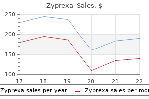
7.5 mg zyprexa saleThe most common malignant lesions of the synovium are metastatic carcinomas medicine quotes buy 2.5 mg zyprexa mastercard, significantly adenocarcinoma of the colon symptoms panic attack zyprexa 5 mg buy with visa, breast and lung. It is unusual for primary malignant bone tumors to prolong into the joint, though they could invade the joint capsule from the delicate tissues. A Ganglion Is a Small Fluid-Filled Cyst A ganglion is a thin-walled, simple cyst containing clear mucinous fluid, which occurs most commonly on the extensor surfaces of the hands and feet, especially the wrist. The cyst arises either from the synovium or from areas of myxoid change in the connective tissue, presumably after trauma. The lesion may be painful and can be readily removed surgically, although a blow with the family Bible was the traditional remedy for a ganglion on the dorsum of the wrist. It is most often seen in association with various types of arthritis, during which the intra-articular stress is increased. The cyst incorporates synovial fluid and microscopically demonstrates a synovial cell lining. Clonal karyotypic abnormalities have been detected in a number of cases of synovial chondromatosis, with diploid or near-diploid complements, chromosome 6 anomalies, rearrangements of 1p22 and 1p13 and additional copies of chromosome 5. Tenosynovial Giant Cell Tumor Is a Benign Neoplasm of Synovial Lining this is the most common neoplasm of the synovium and tendon sheath and occurs in a localized and a diffuse form. Synovial Chondromatosis Features Cartilage Nodules in a Joint Synovial chondromatosis is a benign, self-limited disease by which hyaline cartilage nodules form within the synovium, detach from that construction and float within the synovial fluid, like grains of sand between gears. The persistent irritation produced by these "loose" bodies stimulates the synovium to secrete large quantities of synovial fluid and also causes bleeding within the synovial membrane. Synovial chondromatosis entails the large diarthrodial joints of younger and middleaged males, affecting the knee typically, but additionally the hip, elbow, shoulder, ankle and temporomandibular joints. Patients have pain, stiffness and locking of the joint, with related bloody effusions. Unlike cartilage that detaches from articular surfaces in osteoarthritis, in synovial chondromatosis the fragments of hyaline cartilage are shaped de novo in the synovium. Occasionally, the cartilage nodules, whereas still within the synovium, endure endochondral ossification, during which case the illness known as synovial osteochondromatosis. It occurs principally in younger and middle-aged women (30�50 years) and includes flexor surfaces of the center or index fingers. It involves a single joint, usually in young adults, and is seen equally in men and women. The most typical site is the knee (80%), nevertheless it also happens within the hip, ankle, calcaneocuboid joint, elbow and, less frequently, tendon sheaths of the fingers and toes. Tumors recur in 10%� 20% of circumstances of localized tenosynovial giant cell tumor, in distinction to 40%�50% in the diffuse kind. It might insinuate via joint capsules into gentle tissue and embody nerves and arteries, generally necessitating radical surgical excision. The synovium develops enlarged folds and nodular excrescences, which are brown colored owing to their iron pigment content. The diffuse kind exten- Soft Tissue Tumors Soft tissue tumors are mesenchymal neoplasms that may arise anyplace within the physique however are mostly found within skeletal muscle, fat, fibrous tissue or blood vessels. Tumors of peripheral nerves (see Chapter 32) and other tumors of neuroectodermal differentiation could also be included within the category of soppy tissue tumors. Radiograph of the knee demonstrates confluent erosions of the distal femur and proximal tibia and a delicate tissue mass within the joint. At larger energy, the cellular infiltrate mainly consists of mononuclear histiocytic synoviocytes, lots of which contain brown hemosiderin pigment, and multinucleated big cells. Although delicate tissue tumors could show proof of differentiation towards a selected cell type (fibroblastic, adipocytic, vascular, myoid, and so on. Not all delicate tissue tumors could be readily classified by their line of differentiation. However, many do have attribute and distinctive genomic abnormalities that are diagnostically useful (Table 30-2). Malignant delicate tissue tumors (sarcomas) can metastasize through the bloodstream, often to the lungs or bone. Patients typically die of metastatic disease rather than local invasion at the main tumor website. The ability to distinguish sarcoma from benign mimics is key to prognostication; outcome is based upon each tumor grade and stage. These criteria are mixed with grade and metastatic status for general staging and threat prediction. A group of genetic disorders associated with gentle tissue tumors includes neurofibromatosis kind 1, tuberous sclerosis, Osler-Weber-Rendu disease, Li-Fraumeni syndrome and Gardner syndrome. Burns in childhood produce scars, which in uncommon cases lead to delicate tissue fibroblastic tumors a few years later. Radiation harm can also contribute to the event of sarcomas, in particular angiosarcoma, osteosarcoma or undifferentiated sarcoma, years after publicity. Elongated spindle and stellate cells are arranged haphazardly in a loose myxoid stroma, giving the lesion a "tissue culture�like" look. Rapidly growing tumors are extra likely to be malignant than tumors that develop slowly. Benign tumors are relatively avascular, whereas most malignant ones are hypervascular. Some soft tissue tumors are categorized on the basis of genetic or molecular findings. Most instances occur in young adults who current to medical consideration following the rapid development of the lesion. While nodular fasciitis was lengthy thought to be a posttraumatic reactive condition, the discovery of a recurrent translocation and associated chimeric fusion gene has resulted in a reclassification of this tumor as a type of neoplasia. In addition, cytogenetic abnormalities involving chromosome 15 have been reported in some circumstances. The affected region on chromosome 15 codes for several proteins concerned in tissue repair. Despite these underlying genetic alterations, nodular fasciitis is self-limited and is cured by surgical excision. Fibromatosis Is a Locally Aggressive Proliferation of Fibroblasts Fibromatosis is a regionally invasive, slowly growing mass that will happen virtually anywhere within the physique. Although histologically similar, there are genetic distinctions between superficial and deep "aggressive" variants of fibromatosis. Diabetics, alcoholics and epileptics have an elevated incidence of fibromatosis, as do patients with familial adenomatous polyposis. Microscopically, the lesion is composed of fascicles of bland spindle cells arrayed in long sweeping fascicles in a collagenous stroma.
Zyprexa 5 mg order lineThe development plate controls the longitudinal growth of bones and ultimately determines adult height medicine for uti buy discount zyprexa 5 mg on-line. This part of a short tubular bone demonstrates the first true bone tissue deposited on the skin of the midshaft of the cartilage mannequin together with very early hollowing of the center of the cartilage model to kind mixed spicules of cartilage and bone (primary spongiosa) medicine vs surgery zyprexa 10 mg purchase otc. The epiphysis is separated from the epiphyseal plate by transverse plates of bone that seal the plate so that it grows solely toward the metaphysis. As the calcified cartilage migrates towards the metaphysis, the chondrocytes die, and the lacunae are empty. At the interface of the epiphyseal plate and the metaphysis, osteoclasts bore into the calcified cartilage, accompanied by a capillary loop from the metaphyseal vessels. Osteoblasts follow the osteoclasts and lay down woven bone on the cartilage core, thereby forming the first spongiosum or major trabeculae. The epiphyseal cartilage has ceased to grow, and metaphyseal vessels penetrate the cartilage plate. Viewed longitudinally, the growth plate, continuing from epiphysis to metaphysis, is split into zones. The reserve (resting) zone is provided by epiphyseal arteries and has small chondrocytes and little or no matrix. An additional peripheral zone, generally recognized as the zone of Ranvier, lies instantly underneath the perichondrium. The proliferative zone is the next deeper zone, during which active proliferation of chondrocytes happens both longitudinally and transversely, though the main growth thrust is longitudinal. In a very lively development plate, proliferative zones account for over half the thickness of the growth plate. The hypertrophic zone is next and demonstrates a substantial increase in chondrocyte size. The intercellular matrix is distinguished, and a dense zone, the territorial matrix, surrounds chondrocytes. The zone of calcification is the cartilaginous zone closest to the metaphysis, where the matrix turns into mineralized. Capillaries develop into the calcified cartilage and provides access to osteoclasts, which resorb a lot of the calcified matrix. Residual vertical walls of calcified cartilage act as scaffolding for the deposition of bone. The molecular mechanisms governing endochondral progress are starting to be understood. The misshapen epiphyses seen on radiography mirror incomplete penetration of the secondary centers of ossification of the epiphysis. Morquio Syndrome Features Mucopolysaccharide Deposition in Chondrocytes Many of the mucopolysaccharidoses (see Chapter 6) contain skeletal deformities, attributable to deposition of mucopolysaccharides (glycosaminoglycans) in growing bones. Achondroplasia Is an Inherited Dwarfism Caused by Arrest of the Growth Plate Achondroplasia refers to a syndrome of short-limbed dwarfism and macrocephaly and represents a failure of regular epiphyseal cartilage formation. It is the most typical genetic form of dwarfism (1 in 15,000 stay births) and is inherited as an autosomal dominant trait. The imply grownup peak in achondroplasia is 131 cm (51 inches) in men and 125 cm (49 inches) in girls. Formation of the Metaphysis Is Called Funnelization It happens at the ring of Delacroix, a periosteal cuff of bone surrounding the epiphyseal cartilage. A wave of periosteal osteoclasts resorbs the cortex, in order that a fluted or funnel shape begins to appear. At the identical time, endosteal osteoblastic bone is deposited to maintain tempo with, and offset, a few of the osteoclastic resorption. The Growth Plate Is Normally Obliterated at a Specific Age for Each Bone Closure of the growth plate. In some individuals, a transverse bony plate representing the location of closure could be seen on radiography. The zone of provisional calcification, if present, undergoes endochondral ossification, however at a significantly decreased rate. A transverse bar of bone often seals off the growth plate, thus preventing further bone formation and inflicting dwarfism. Because intramembranous ossification is undisturbed, the periosteum functions usually and the bones turn into very brief and thick. Thyroid hormone plays a task in regulating chondrocytes, osteoblasts and osteoclasts via production of cytokines and different factors involved in bone improvement and development. Linear growth is severely impaired, resulting in dwarfism, with limbs disproportionately brief in relation to the trunk. There is a delay in closure of the epiphyses, as nicely as radiologic stippling of these zones. Instead, maturation of the hypertrophied zone is retarded, and the zone of proliferative cartilage is slender. In achondroplasia, the epiphyseal plate is lowered in thickness, and the zones of proliferating cartilage are attenuated. Osteoclastic exercise is inconspicuous, and the interface between the plate and the metaphysis is usually sealed by transverse bars of bone that stop additional endochondral ossification. Furthermore, the basement membrane of capillaries is broken by this situation and widespread capillary bleeding is frequent. An arteriovenous malformation may trigger one progress plate to develop sooner than its counterpart, as may fractures and tumors close to the expansion plate. Asymmetric Cartilage Growth Causes Spinal Disorders and Tumors Asymmetric cartilage progress, similar to happens in patients with knock-knees and bowed legs, develops when one part of the expansion plate, either medial or lateral, grows sooner than the opposite. Most instances are hereditary, but mechanical forces such as trauma near the expansion plate might stimulate one side to grow sooner or in an uneven fashion. Aside from the beauty look, these situations could require correction to forestall future incongruity, eventual loss of articular cartilage and joint destruction. Death of infants with this extreme variant is attributable to marked anemia, cranial nerve entrapment, hydrocephalus and an infection. A more benign Scoliosis and Kyphosis Scoliosis is an abnormal lateral curvature of the spine, often affecting adolescent ladies. The illness is brought on by mutations in genes that govern osteoclast formation or perform. The commonest mutations trigger defects in bone acidification, which is critical for osteoclastic bone resorption. Other mutations that cause osteopetrosis involve transcription elements or cytokines needed for the osteoclast differentiation. These bones are extraordinarily radiopaque and weigh two to thrice greater than regular bone. The mineralized cartilage can additionally be weak and friable, in order that the bones in osteopetrosis fracture simply. Grossly, bones in osteopetrosis are widened in the metaphysis and diaphysis, causing the characteristic "Erlenmeyer flask" deformity.
2.5 mg zyprexa overnight deliveryOther causes embrace elevated residual urine volume because excessive levels of progesterone make bladder musculature flaccid and fewer able to treatment of schizophrenia order zyprexa 7.5 mg without a prescription expel urine treatment bladder infection zyprexa 7.5 mg buy cheap. Subsequent addition of sterile urine from the kidneys dilutes any micro organism which will have gained access to the bladder. Diabetic glycosuria additionally facilitates infection by providing a rich bacterial progress medium. Increased intravesicular strain during micturition occludes the distal ureteral lumen and prevents urinary reflux. An anatomic abnormality, a short passage of the ureter throughout the bladder wall, causes the ureter to insert extra perpendicularly to the bladder mucosal floor. As a outcome, rather than occluding the lumen, micturition will increase intravesicular pressure and pushes urine into the patent ureter. The convexity of the straightforward papillae of central calyces blocks reflux urine from entering. However, if pressure is prolonged, as in obstructive uropathy, even easy papillae are eventually vulnerable to retrograde entry of urine. Anatomic features of the bladder and kidney in pyelonephritis attributable to ureterovesical reflux. On micturition, the elevated intravesicular pressure compresses the flap against the bladder wall, occluding the lumen. For instance, in bacterial endocarditis, gram-positive organisms, corresponding to staphylococci, can unfold from an contaminated valve and establish an infection in the kidney. In extreme cases of acute pyelonephritis, necrosis of the papillary tips could happen. Renal parenchyma, notably the cortex, usually reveals extensive focal destruction by irritation, although vessels and glomeruli often are preferentially preserved. Whether reflux with out an infection can produce continual pyelonephritis is controversial. Differentiating higher from decrease urinary tract infections clinically is commonly tough, however leukocyte casts in the urine recommend pyelonephritis. The bisected kidney shows a dilated renal pelvis and dilated calyces secondary to urinary tract obstruction. The papillae are all necrotic and appear as sharply demarcated, ragged, yellowish areas. An in depth infiltrate of neutrophils is present within the collecting tubules and interstitial tissue. Many ailments cause persistent injury to the tubulointerstitial compartment and induce chronic interstitial inflammation, interstitial fibrosis and tubular atrophy. Thus, continual pyelonephritis is considered one of many causes of a sample of injury known as chronic tubulointerstitial nephritis. Vesicoureteral reflux causes an infection of the peripheral compound papillae and, subsequently, scars in the poles of the kidney. Obstruction of the urinary tract leads to high-pressure backflow of urine, which causes an infection of all papillae, diffuse scarring of the kidney and thinning of the cortex. In obstructive uropathy, all of the calyces and the renal pelvis are dilated, and the parenchyma is uniformly thinned. In circumstances related to vesicoureteral reflux, the calyces on the poles of the kidney are preferentially expanded and are related to overlying discrete, coarse scars that indent the renal floor. The scars have atrophic dilated tubules surrounded by interstitial fibrosis and chronic inflammatory infiltrates. The most characteristic (but not specific) tubular change is severe epithelial atrophy, with diffuse, eosinophilic, hyaline casts. Such tubules are "pinched-off" spherical segments, resembling colloid-containing thyroid follicles. This pattern, called thyroidization, outcomes from breakup of tubules and residual segments forming spherules. Its name derives from the yellow gross appearance of nodular renal lesions, attributable to numerous lipid-laden foamy macrophages (xanthoma cells). Analgesic Nephropathy Results from Chronic Overconsumption of Phenacetin Patients with analgesic nephropathy sometimes have taken greater than 2 kg of analgesics, usually in combinations, such as aspirin and phenacetin, or aspirin and acetaminophen. Phenacetin most frequently results in nephropathy and is banned in lots of nations, including the United States. Possibilities include direct nephrotoxicity, ischemic harm because of druginduced vascular changes or both. Many tubules contain eosinophilic hyaline casts resembling the colloid of thyroid follicles (so-called thyroidization). The earliest histologic abnormality is a particular homogeneous thickening of capillary partitions just beneath the transitional epithelium of the urinary tract. The lesion is characterized by a granulomatous response, filled with foamy histiocytes. Early parenchymal changes are confined to papillae and the inside medulla, they usually encompass focal basement membrane thickening of tubules and capillaries, interstitial fibrosis and focal coagulative necrosis. Necrotic areas ultimately become confluent, first affecting the corticomedullary junction and then the accumulating ducts. Eventually, the entire papilla becomes necrotic (papillary necrosis), typically remaining in place as an amorphous mass. Papillae may stay partly attached at the demarcation zone or be utterly sloughed. There is secondary tubular atrophy, interstitial fibrosis and continual irritation within the overlying cortex. Neutrophils are rare; their presence ought to elevate suspicion of pyelonephritis or hematogenous bacterial an infection. Sloughing of necrotic papillary ideas into the renal pelvis might lead to colic as they pass through the ureters. The immunogen could be the drug itself, the drug sure to certain tissue components, a drug metabolite or a tissue part altered by the drug. There is interstitial edema and infiltration by mononuclear leukocytes, with admixed eosinophils. Urinalysis exhibits erythrocytes, leukocytes (including eosinophils) and sometimes leukocyte casts. Most patients recover fully inside several weeks or months if the offending drug is discontinued. Light-Chain Cast Nephropathy May Complicate Multiple Myeloma Light-chain solid nephropathy is renal harm attributable to monoclonal immunoglobulin light chains in the urine. However, at the acidic pH typical of urine, these mild chains kind casts by binding to Tamm-Horsfall glycoproteins which are secreted by distal tubular epithelial cells.
2.5 mg zyprexa with mastercardAccumulation of neutrophils in glomerular capillaries portends impending rejection treatment advocacy center zyprexa 20 mg buy without prescription. Endothelial cell adjustments are adopted by platelet thrombi and later by fibrin thrombi treatment episode data set discount 20 mg zyprexa visa. Neutrophils or mononuclear leukocytes are elevated in peritubular and glomerular capillaries, and in tubules. Complement activation merchandise, especially C4d, localize constantly to the walls of peritubular and glomerular capillaries. The most extreme, however least frequent, sample of acute antibody-mediated rejection involves necrotizing arteritis with fibrinoid necrosis of the media. Preformed antibody in opposition to recipient antigens causes a direct in situ reaction, with hemorrhage developing as a result of vascular necrosis. Staining of peritubular and glomerular capillaries with an anti-C4d antibody exhibiting proof of complement activation by antibodies directed in opposition to donor antigens on endothelial cells. Acute antibodymediated necrotizing acute vasculitis in an interlobular artery with intensive fibrinoid necrosis of the muscularis. The vascular and interstitial infiltrates of mononuclear leukocytes indicate concurrent acute cellular rejection. If necrotizing arteritis develops, fewer than 30% of grafts survive 1 year, even with aggressive immunosuppression. It is characterized by infiltration of the interstitium, tubules, arteries, arterioles or glomeruli by T lymphocytes and macrophages. Nuclei of infiltrating lymphocytes vary in dimension and form as a end result of the cells are at various levels of activation and embrace immunoblasts (see Chapter 26). Involvement of tubules (tubulitis) is manifested by lymphocytes crossing tubular basement membranes and mendacity between tubular epithelial cells. Arterial involvement by cellular rejection entails T lymphocytes and monocytes traversing the endothelium, increasing the intima with mononuclear leukocytes (endarteritis). Glomerular infiltration by mononuclear leukocytes with obliteration of capillary lumens causes acute transplant glomerulitis). Renal transplants with tubulitis but not endarteritis have an 80% likelihood of 1-year graft survival, compared with 60% for allografts with endarteritis. Chronic transplant arteriopathy impacts arteries of all sizes, together with the main renal artery. Inflammation is absent-or is much much less distinguished than in active acute cellmediated intimal arteritis (compare to . Foam cells may be conspicuous, and the inner elastic lamina could also be interrupted. Ischemia due to arterial and capillary narrowing might result in tubular atrophy and interstitial fibrosis. Acute tubulointerstitial mobile rejection with tubulitis indicated by lymphocytes on the epithelial aspect of the basement membrane (periodic acid�Schiff stain). Acute cellular vascular rejection with endarteritis indicated by mononuclear leukocytes infiltrating the intima of an arcuate artery. The lumen of this mediumsized artery is occluded by a thickened intima, which contains a number of inflammatory cells. The frequency and significance of recurrence differ among different sorts of glomerular disease (Table 22-15). Unfortunately, each medicine are nephrotoxic and can injure each kidney allografts and native kidneys of patients given these medicine for other reasons. The most characteristic renal lesion is an arteriolopathy that begins with easy muscle cell degeneration and necrosis. The destroyed arteriolar muscle cells are changed by acidophilic hyaline material. In fulminant cases, vascular lesions resemble full-blown thrombotic microangiopathy, with circumferential fibrinoid arteriolar necrosis. In chronic toxicity, there are zones of interstitial fibrosis and tubular atrophy ("striped fibrosis"). Intranuclear viral inclusions in tubular cells may suggest this risk, and immunochemical staining can affirm the diagnosis. Tumor size, which has been used to separate adenomas from carcinomas, is problematic as a outcome of all carcinomas begin out as small lesions. Neoplasms smaller than 5 mm with papillary or tubulopapillary growth patterns could be thought-about adenomas. Papillary renal adenomas occur extra often with advancing age and are incidental post-mortem findings in 40% of patients older than 70. They are plump, with abundant, finely granular, acidophilic cytoplasm and spherical nuclei that lack atypia. The distinctive appearance of the tumor is because of plentiful mitochondria in the cytoplasm. Oncocytomas are sometimes mahogany-brown as a result of mitochondrial lipochrome pigments. Renal medullary fibromas are incidental findings in as many as half of all grownup autopsies. These lesions are mixtures of well-differentiated adipose tissue, easy muscle and thick-walled vessels. The lesions vary from smaller than 1 cm to bigger than 15 cm and are composed of spindle cells of fibroblastic or myofibroblastic lineage. Tumor margins are normally irregular, with bands of cells interdigitating with adjacent renal parenchyma. Mesoblastic nephromas may recur if a few of these tongues of tumor tissue are left behind after surgical resection. Its incidence is 1 in 10,000, making it the most typical stomach stable tumor in kids. Another risk is that one other closely linked gene, such as the H19 gene, expressed solely by the maternal allele, is a tumor suppressor or regulates imprinting in the area. Nephrogenic rests (small foci of persistent primitive blastemal cells) are discovered in the kidneys of all kids with syndromic Wilms tumors and in 1/3 of sporadic cases. Most Wilms tumors contain all three parts in various proportions, however only two parts or even just one could additionally be present every so often. This Wilms tumor reveals extremely cellular areas composed of undifferentiated blastema (B), loose stroma (S) containing undifferentiated mesenchymal cells and immature tubules (T). The tumor stroma incorporates spindle cells, that are largely undifferentiated but could show easy muscle or fibroblast differentiation. Skeletal muscle is the most typical heterotopic stromal factor, although bone, cartilage, fat or neural tissue may not often be encountered. They occur in 1 in 10,000 kids, usually 1�3 years old, and 98% present before age 10. Several histologic and medical parameters are used to predict the habits of these tumors, with various success.
Buy zyprexa 2.5 mg with amexThe safe and efficacious use of these brokers has been outlined in evidence-based guidelines to give guidance to clinicians treating complex ache patients symptoms 9 dpo zyprexa 5 mg best. This paved the way in which for the first utility of this idea in humans and publication in 1979 of a series of eight patients on the Mayo Clinic with metastatic cancer who have been handled with intrathecally delivered opioids treatment modality definition buy cheap zyprexa 5 mg on line. The danger of demise appeared attributable to identifiable causes such as adjuvant medication, coexisting diseases, and improper drug alternatives but to not gear malfunction. The suggestions included increased monitoring, lowered initial starting doses of opioids, and therapy of coexisting diseases. More widespread issues include catheter migration, catheter fracture or kinking, superficial infection, postdural puncture headache, and nerve irritation. Other frequent problems embrace drug reactions including peripheral edema, intrathecal granuloma, low testosterone, and urinary retention. Of notice, acute baclofen withdrawal can cause severe reactions and multisystem organ failure leading to dying. The use of low doses of intrathecal drugs and drug combinations can result in improved operate, decreased pain, and improved high quality of life. The improvement of recent medicine and methods for targeted drug delivery will more than likely lead us to new frontiers including the treatment of problematic points corresponding to melancholy, epilepsy, amyotrophic lateral sclerosis, and other nervous system disorders. Hayek S, Deer T, Pope J, Panchal S, and Patel V (2011) Intrathecal therapy for most cancers and non-cancer ache. Royster E and Crumbley K (2011) Initial experience with implanted peripheral nerve stimulation for the remedy of refractory cephalgia. Simopolous T, Bajwa Z, Lantz G, Lee S, and Burstein R (2010) Implanted auricular nerve stimulator for the remedy of refractory chronic migraine. The neuromuscular problems comprise a variety of illness states that affect the decrease motor and sensory neurons, the spinal (motor and sensory) roots, peripheral nerves, neuromuscular junctions, and muscle tissue. The authors describe the scientific manifestations related to problems affecting these components of the peripheral nervous system and touch upon their underlying causes. Weakness is characterized by the lack of strength and may be described as gentle, average, or severe. Alternatively, it might be measured by the Medical Research Council scale during which power is rated from 5 (normal) to zero (no motion at all). Fasciculations are spontaneous seen twitches in a muscle ensuing from the contractions of most of the individual muscle fibers belonging to an electrically excitable motor unit. For instance, degeneration and lack of motor neurons in the best hypoglossal nucleus in the medulla of the brainstem would be anticipated to trigger weak spot, atrophy, and fasciculations of the proper aspect of the tongue. A comparable process affecting motor neurons in the best cervical spinal wire would cause lower motor neuron indicators in the best hand. Note the motor neuron, its axon, and lots of muscle fibers which are innervated by it. There are small groups of angulated atrophic fibers (arrows) that have lost their innervation. When Betz cells are lost, a group of medical findings emerge which are referred to as upper motor neuron signs. These comprise increased muscle tone or spasticity, elevated reflex exercise or hyperreflexia, and pathological reflexes, such as the Babinski sign. Causes of Motor Neuron Disorders There are two main categories of motor neuron problems: genetically decided and acquired. There can be an grownup In some clinical settings, degeneration of lower motor neurons is accompanied by lack of cortical motor neurons. These latter cells are additionally referred to as Betz cells, and they project closely myelinated fibers to connect with decrease motor neurons of the brainstem and spinal twine. Degeneration or loss of peripheral sensory neurons positioned within the spinal sensory dorsal root ganglia result in some combination of the following signs and symptoms: sensory impairment, disappearance of tendon reflexes, impaired coordination as a result of defective proprioception, and pain and paresthesias. Disorders that affect these sensory neurons completely are known as sensory neuronopathies, and the ensuing scientific picture is purely sensory (sensory symptoms and indicators, including impairment in proprioception that may lead to pseudoathetosis of affected arms and legs) whereas motor function is preserved. Causes of Sensory Neuron Disorders As noted for the motor neuron problems, there are additionally two major categories of sensory neuron disorders: genetically decided and acquired. A typical presentation for an acquired sensory neuronopathy would possibly comprise the subacute to chronic evolution (over weeks to months) of tingling paresthesias and ache within the feet, lack of manual dexterity, and poor steadiness with frequent falls. Also shown is the peripheral Damage to a single nerve root or to many roots will lead to a set of signs and signs referable to both a particular injured root (referred to as a radiculopathy) or to a quantity of roots (in which case the dysfunction is designated a polyradiculopathy). In the cervical area, the most common website of radiculopathy is either C6 or C7 and in the lumbosacral region, the L5 or S1 roots are most commonly affected. The symptoms of root involvement are numbness, pain, and weakness in both the arm or leg, and the indicators comprise loss of sensation within the dermatome (the sensory territory of the affected root), reduction or lack of reflex activity subserved by the nerve root and its respective reflex arc in question, and weak point in the myotome (the motor territory innervated by the basis in question). For example, a affected person with a C7 radiculopathy would present with ache within the shoulder and scapular area, numbness in the center finger (the territory of the C7 dermatome), weak spot within the triceps and wrist flexor (muscles which have innervation from the C7 myotome), and loss of the triceps reflex (caused by interruption of sensory afferents within the C7 nerve root). When the illness course of affects thoracic roots, the major manifestation is ache and numbness within the dermatomes of the affected nerve roots, normally similar to a area on the chest wall, extending from the sternum alongside the entrance of the chest to the facet and then additional toward the spine. Disorders of Peripheral Nerve Clinical Features There are three major scientific presentations of peripheral nerve illness: focal, multifocal, and diffuse. The focal category comprises mononeuropathy, characterised by numbness, sensory loss, pain, and weak point in the territory of an affected nerve. Multifocal peripheral nerve disease is characterised by multiple mononeuropathies wherein symptoms and signs happen within the distribution of two or more nerves. The diffuse disorder, polyneuropathy, is normally characterised by signs and indicators referable to dysfunction of each sensory and motor fibers, with findings extensively and symmetrically distributed, affecting the decrease and the upper extremities. In some cases, the disorders inflicting polyneuropathy target sensory, autonomic, or motor fibers selectively. The medical manifestations of polyneuropathy comprise signs of numbness and tingling in addition to weak point. Typically, distal parts of the extremities are affected earlier than the more proximal, and the legs before the arms. Accordingly, within the early phases of the disease course of, the bodily examination findings are loss of sensation in the toes and attenuated tendon reflexes on the ankles. At that early stage, motor findings may be minimal or restricted to mild atrophy and weak spot of intrinsic foot muscular tissues. With continued exercise of the illness process, sensory loss could unfold extra proximally to the toes and ankles and probably to more rostral ranges. At a later stage of the disease, the arms can also become concerned with sensory loss and weak point of hand intrinsic muscle tissue (those that comprise the thenar (base of the thumb) and hypothenar (base of the little finger) eminences). A much less frequent medical state of affairs for the presentation of a Causes of Nerve Root Disorders the commonest causes of radiculopathy are structural: nerve root compression by a herniated nucleus pulposus or by osteophytes complicating spondylotic arthropathy. Causes of Peripheral Nerve Disorders the mononeuropathies are most often caused by compression or entrapment, with median mononeuropathy on the wrist or peroneal mononeuropathy on the fibular head being widespread examples. The causes of polyneuropathy are quite a few, including diabetic, immunological, infectious, toxic, paraneoplastic, and heredofamilial. The most common postsynaptic dysfunction, myasthenia gravis, presents with weak point affecting all skeletal muscles, especially those which would possibly be cranial nerve innervated, particularly ocular muscular tissues. Typically, early symptoms embody lid droop (ptosis) and double vision (diplopia) followed in some patients by problem in chewing, swallowing (dysphagia), and speaking (dysarthria) and possibly generalized limb weak point. In a small percentage of sufferers, respiratory muscle weak point may develop, during which case the diploma of severity of the myasthenia reaches the crisis stage, requiring acute medical intervention and hospitalization in the intensive care unit.

7.5 mg zyprexa with visaPhotomicrograph displaying irregular cords and nests of invasive ductal carcinoma cells invading stroma medicine zanaflex order zyprexa 20 mg without prescription. In distinction to invasive ductal carcinoma medications of the same type are known as 7.5 mg zyprexa order with mastercard, the cells of lobular carcinoma tend to kind single strands that invade between collagen fibers in a diffuse pattern. If a special-type part makes up over 50% of the tumor, the tumor is considered blended. Specific genetic lesions or alterations are related to a specific histologic sort or grade in some instances. Tubular carcinomas are well-defined stellate lots whose mobile composition is kind of entirely open and angulated tubules, lined by a single layer of mildly atypical epithelial cells. Tubular carcinomas share some patterns of karyotypic modifications with other tumor types (Table 25-1). Lymph node metastases are uncommon, and patients with tubular carcinomas have an excellent prognosis. Invasive Lobular Carcinoma Invasive lobular carcinoma is the second most typical type of invasive breast cancer, accounting for 5%�15% of all invasive carcinomas. Because stromal desmoplasia and fibrosis may be minimal, sufferers often have clinically silent illness grossly and by mammography, or might current with a poorly defined thickening of the breast. Lobular cancers characteristically show dishesive malignant epithelial cells that infiltrate the stroma diffusely. E-cadherin expression is often low or absent, reflecting biallelic lack of the tumor suppressor gene that encodes this protein. Patterns of genetic adjustments in invasive lobular carcinoma differ from these in ductal carcinomas (Table 25-1). Ovary displaying metastatic lobular carcinoma characterized by dyshesive cells with eccentric nuclei and intracytoplasmic lumens. Open and angulated malignant glands are dispersed between regular lobules and present extension into fats. A single layer of epithelium lines the tubules, and myoepithelial cells are absent. The malignant cells are pleomorphic and develop in strong sheets, forming a blunt margin. Sponge-like pattern of empty areas containing glands and small clusters of malignant epithelium. Cartilaginous and osseus matrix in a metaplastic carcinoma with heterologous parts. Mucinous Carcinoma Patients with mucinous carcinoma are usually older than those with different tumor sorts. These tumors, which make up 1%�6% of breast cancers, are nicely circumscribed, with a gelatinous texture. Low-grade malignant epithelial cells kind acini, nests or trabeculae, which seem to float in swimming pools of extracellular mucin. Pure mucinous carcinomas show little genomic instability or recurrent amplifications. Carcinomas with Medullary Features Classic medullary carcinomas are exceptionally uncommon, though different forms of carcinoma might present medullary options. In these tumors, malignant epithelial nests or acini are surrounded by a transparent space. Some locally superior tumors are staged T4, based mostly on pores and skin or chest wall invasion, regardless of tumor measurement. Arm edema and pain can also happen, in all probability due to lymphatic obstruction by tumor. Metaplastic Carcinoma Metaplastic carcinomas are heterogeneous tumors with malignant spindle cells, squamous cell carcinoma or heterologous elements, similar to bone or cartilage. Adenocarcinoma could additionally be absent, however cytokeratin immunostains are at least focally current. These tumors typically cluster with the basal molecular subgroup on gene expression profiling (see below). Low-grade, fibromatosis-like, metaplastic carcinoma and low-grade adenosquamous metaplastic carcinoma are associated with a favorable end result. Other metaplastic subtypes reply poorly to adjuvant chemotherapy and fare worse than other forms of triple-negative breast cancer. Lymph Node Status the presence or absence of axillary lymph node metastases is a key prognostic indicator for patients with breast most cancers and requires pathologic evaluation of surgically resected lymph nodes. This process requires injection of a dye and radioactive isotope and involves intraoperative lymphatic mapping of the draining or "sentinel" lymph node, the node more than likely to include breast cancer metastases. Immunohistochemical staining may help to determine cytokeratin-positive epithelial cells that is in all probability not seen in any other case. The actual impact on prognosis of small metastases is small in contrast with node-negative women. Breast cancers metastasize to bone, which is the place metastatic disease presents in 25% of circumstances. The Nottingham grading system, also referred to as the modified Bloom and Richardson methodology, is most generally used. It combines scores for tubule formation, nuclear pleomorphism and mitotic count into a ultimate grade of 1, 2 or 3 for low-, intermediate- and high-grade carcinomas, respectively. Patients with grade 1 tumors have considerably better survival than those with grade 2 or grade 3 tumors. Low-grade invasive carcinoma showing good tubule formation, gentle nuclear pleomorphism and inconspicuous mitoses. Moderately differentiated carcinoma with less tubule formation, reasonable nuclear pleomorphism and variably distinguished mitoses. Poorly differentiated carcinoma exhibiting absent tubule formation, marked nuclear pleomorphism and frequent mitotic figures. Proliferative index and ploidy: Tumors with excessive proliferative indices have worse prognoses. Several parameters are used to assess proliferation in breast cancers, together with (1) mitotic index, assessed histologically; (2) the proportion of cells in S phase of the cell cycle by move cytometry; (3) immunohistochemical staining for proteins (Ki67) expressed by actively proliferating cells. Hormone receptor positivity is outlined as higher than or equal to 1% staining tumor cells. The biggest value of assessing hormone receptor standing in breast most cancers is its predictive capability. Immunohistochemistry detects cell membrane expression of the protein, and in situ hybridization identifies gene amplification. Strong nuclear positivity for estrogen receptor on this reasonably differentiated invasive ductal carcinoma (immunohistochemical stain). Notably, a lot of the prognostic influence of multigene predictor signatures (discussed below) comes from proliferation genes. Response to neoadjuvant therapy: In sufferers who obtain systemic therapy before surgical procedure (neoadjuvant therapy), the response to the therapy is a powerful prognostic issue. Poorly differentiated tumors with high proliferation indices are more likely to respond to neoadjuvant therapy than low-grade cancers. A pathologically complete response happens in 10%�30% of patients, little or no response in 10%�15% of sufferers and partial response in the the rest.
Diseases - Hypertonic gingivitus
- Hypercholesterolemia due to LDL receptor deficiency
- Acute monocytic leukemia
- Dysostosis acral with facial and genital abnormalities
- Familial cold autoinflamatory syndrome (FCAS)
- Chondrysplasia punctata, humero-metacarpal type
- HIV
- D-plus hemolytic uremic syndrome
Cheap zyprexa 20 mg with visaIs the affected person unable to shut his or her eyes utterly when requested to do so chi infra treatment generic zyprexa 7.5 mg without a prescription, indicating higher facial muscle weak spot Finally symptoms nasal polyps purchase 10 mg zyprexa, if the affected person complains of muscle stiffness, the doctor ought to attempt to elicit myotonia. Facial myotonia can be noticed after repeated forceful voluntary eye closure. Once the myopathy is advanced, and the muscular tissues are extremely weak and atrophic, stretch reflexes might become hypoactive or unelicitable. Patterns of Weakness Once the muscles have been inspected, examined for power, and useful exercise has been noticed, an attempt must be made to place the affected person in one of the patterns of muscle weakness that may occur in myopathic disorders. This sample of weak point could be seen in lots of hereditary and bought myopathies and due to this fact is the least particular in arriving at a selected analysis. The pattern of distal weak point in the higher or lower extremities (anterior or posterior compartment muscle groups) (Table 3). Selective weakness and atrophy in distal extremity muscular tissues is more typically a feature of neuropathies however is rare because of a major muscle disease. When this sample of weak spot is determined to be due to a myopathic rather than a neuropathic disorder, a diagnosis of distal myopathy is acceptable. The sample of distal upper extremity weak point within the distal forearm muscle tissue (wrist and finger flexors) and proximal lower extremity weakness involving the knee extensors (quadriceps). In addition, the weak spot is usually asymmetrical between the two sides, which is uncommon in most myopathies. Rarely this sample can be seen in myotonic dystrophy but is symmetrical and related to grip myotonia. The combination of ptosis, ophthalmoplegia with out diplopia, and pharyngeal weakness should recommend the diagnosis of oculopharyngeal dystrophy, especially if the onset is in middle age or later. When static, ptosis, and ophthalmoplegia without distinguished pharyngeal involvement is a trademark of most of the mitochondrial myopathies. Fluctuating ptosis and ophthalmoplegia with diplopia are typical of neuromuscular junction defects. Ptosis and facial weak point without ophthalmoplegia or pharyngeal weakness is a standard function of myotonic dystrophy and fascioscapulohumeral dystrophy. Patients with ocular or pharyngeal involvement can also have the everyday pattern of limb-girdle weakness. Prominent neck extensor weak point: Some myopathic circumstances have a dramatic degree of weak point of the neck extensor muscles. Significant neck extensor weak point happens within the setting of a myopathy that also has one of many beforehand outlined phenotypic patterns. For instance, a patient with a limb-girdle sample of weak point may have vital neck extensor involvement. However, sometimes isolated neck extensor myopathy might happen as a definite muscle disorder. Bulbar dysfunction in limb-girdle muscular dystrophy 1A (myotilin) results in dysarthria. Other neuromuscular ailments also can current with certainly one of these weak point patterns. Ocular, pharyngeal, and proximal limb weak point is a frequent function of neuromuscular junction transmission problems such as myasthenia gravis. However, these sufferers may even have diplopia, weakness that fluctuates, and extra laboratory options that ought to result in the right prognosis. Associated Organ Involvement or Systemic Illness Involvement of organs or tissues aside from muscle can present further diagnostic clues. Hepatomegaly can happen in sarcoidosis and in the myopathies associated with deficiencies of acid maltase, debranching enzyme, and carnitine. Intrinsic pulmonary involvement may be seen in a few of the inflammatory myopathies and sarcoidosis. Another spontaneous electrical phenomenon, the complex repetitive discharge, is often the result of nerve disease but is sometimes noticed in myopathies. It is due to muscle fiber ephaptic transmission, which is cross-talk amongst adjoining injured muscle fibers. A fasciculation represents a spontaneous discharge of a motor unit, which is a group of muscle fibers of the identical histochemical type under the control of a single anterior horn cell and thus innervated by the same axon. Electrically, a fasciculation is a large-amplitude potential that consists of the simultaneous involuntary depolarization of a group of muscle fibers. Activity with movement When the patient voluntarily contracts the muscle, individual motor unit motion potentials are assessed. In a myopathy, multiple small motor units are recruited with only minimal voluntary effort. Spontaneous rhythmic discharges of a single muscle fiber, known as fibrillation or constructive sharp waves, occur when there was a disconnection between the nerve and the muscle it innervates. Fibrillations are seen most often in nerve ailments however can occur in active myopathies. For instance, if a muscle fiber turns into injured due to an inflammatory myopathy, it can lose its connection with the nerve terminal and fibrillate. The finding of fibrillation potentials at relaxation in a patient suspected of a myopathy normally signifies lively, ongoing muscle fiber degeneration. Myotonia is one other abnormal spontaneous discharge of muscle that happens in some myopathies, corresponding to myotonic dystrophy, myotonia congenita, periodic paralysis, and acid maltase deficiency. It represents repeated muscle fiber depolarization as a result of an irritable muscle membrane. Muscle Biopsy A muscle specimen may be obtained via either an open or a closed (needle or punch) biopsy process. Most centers perform primarily open muscle biopsies, however this is in giant part determined by the experience of the histology laboratory during which the tissue is processed. Not all laboratories are in a position to course of the tissue adequately for the entire essential research from a small punch biopsy specimen. Occasionally, the muscle to biopsy can be guided by abnormalities on an imaging process, such as muscle ultrasound, computed tomography, or magnetic resonance imaging. In most instances, gentle microscopic observations are sufficient to make a pathological diagnosis. Muscle tissue examined underneath gentle microscopy is primarily performed using frozen specimens. The tissue is examined for muscle fiber size, form, fiber type distribution, and the presence of fiber degeneration (necrosis) or regeneration. Type 1 fibers (slow-twitch, fatigueresistant, and oxidative metabolism) stain lightly at alkaline and darkly at acidic pHs. Type 2 fibers (fast-twitch, fatigueprone, and glycolytic metabolism) stain darkly at alkaline and flippantly at acidic pHs.
Buy 20 mg zyprexa otcPatients present with hypercalcemia medications you cant take while breastfeeding 20 mg zyprexa order free shipping, hypophosphatemia 25 medications to know for nclex order 7.5 mg zyprexa fast delivery, nephrolithiasis and bone illness. In half of sufferers, one gland may be noticeably larger than the others, which can complicate the excellence from adenoma. In hyperplastic glands, the traditional glandular adipose tissue is replaced by hyperplastic chief cells arranged in sheets or trabecular or follicular patterns. An necessary characteristic that distinguishes hyperplasia from adenoma is that hyperplasias lack mobile pleomorphism. Parathyroid Carcinomas Account for 1% of Hyperparathyroidism Parathyroid carcinomas are rare. They are usually functioning tumors, and most sufferers present with symptoms of hyperparathyroidism. Hypercalcemia in these patients is commonly severe, with serum calciums in excess of 14 mg/dL. Parathyroid Adenomas Account for Most Cases of Hyperparathyroidism Solitary parathyroid adenomas trigger 85% of primary hyperparathyroidism. Neck radiation and hereditary syndromes with histories of parathyroid adenomas are risk factors. Other syndromes of increased threat embrace hyperparathyroidism�jaw tumor syndrome and familial isolated hyperparathyroidism. The tumor consists of sheets of neoplastic chief cells and is separated from regular parenchyma by a thin capsule (arrows). This tumor suppressor gene, at chromosome 1q25-31, is liable for the hyperparathyroidism�jaw tumor syndrome. It encodes parafibromin, expression of which is low or absent in hyperparathyroid� jaw tumor syndrome and in carcinomas. Most carcinomas present trabecular cell arrangements, with significant mitotic exercise and thick fibrous bands. The cellular atypia that occurs typically in parathyroid adenomas is rare in carcinomas. After surgical removing, local recurrence is frequent: about 1/3 of sufferers develop metastases to regional lymph nodes, lungs, liver and bone. If deadly, death is most often due to hyperparathyroidism rather than carcinomatosis. The normal adipose tissue of the gland has been changed by sheets and trabeculae of hyperplastic chief cells. These sufferers current with bone pain, bone cysts, pathologic fractures and localized bone swellings (brown tumors, epulis of the jaw). Nephrocalcinosis, observed radiologically as diffuse renal calcification, may happen (see Chapter 22). Peripheral neuropathy with skeletal muscle type 2 fiber atrophy results in weak point. Hypercalcemia may also cause constipation and continual pancreatitis by means not properly understood. Major pathogenetic pathways leading to clinical main and secondary hyperparathyroidism. The parathyroids in secondary hyperplasia resemble these in major chief cell hyperplasia. Treatment is surgical removing of the enlarged glands, with or without reimplantation. Tertiary hyperparathyroidism is autonomous parathyroid hyperplasia following long-standing secondary hyperplasia because of renal failure. In such instances, parathyroid hyperplasia may not regress after renal transplantation, and parathyroidectomy is required. Two thirds of patients with long-standing uremia have monoclonal hyperplastic parathyroid proliferations. Ectopic adrenal tissue can happen in plenty of locations outdoors the gland, for the rationale that cells migrate alongside the gonads. Common places embody the retroperitoneum, the broad ligament close to the ovary, close to the epididymis, the kidney and the liver. Small nodules of adrenocortical cells typically occur within the fibroadipose tissue that surrounds the adrenal gland. Congenital Adrenal Hypoplasia this condition is kind of rare and will accompany renal agenesis. It has been reported in association with toddler demise syndrome and with hypogonadotrophic hypogonadism in adolescents. The cortex arises from celomic mesenchymal cells near the urogenital ridge and secretes steroid hormones corresponding to aldosterone, cortisol and testosterone. Adult adrenal glands are pyramidal organs above every kidney, discovered anteriorly in the retroperitoneum. The adrenal glands secrete steroid hormones or corticosteroids, regulate metabolism and immune system function and help in responses to stress. Grossly, and on reduce part, the cortex has a attribute yellow color because of lipid deposits. Their cut surfaces are soft, tan to brown and either diffusely enlarged or nodular. In most instances, the zona glomerulosa is also hyperplastic, however not to the extent of the opposite zones, particularly the zona fasciculata. Ectopic adrenal tissues or nodules could also be additionally hyperplastic, and if stimulation persists, adenomas can develop. Electron microscopy exhibits that adrenal cortical cells have plentiful easy endoplasmic reticulum and heaps of mitochondria with lamellar cristae, which is common for steroid-producing cells. There, aldosterone manufacturing is stimulated by angiotensin and potassium and inhibited by atrial natriuretic peptide and somatostatin. The zona glomerulosa makes up 15% of the cortex, with vague spherical nests of cells with dark-staining nuclei and moderate numbers of cytoplasmic fat droplets. The zona fasciculata makes up 75% of the cortex and produces glucocorticoids, corresponding to cortisol. This zone contains radial cords of larger cells, every with a small nucleus and a large, foamy, clear cytoplasm, representing saved lipid. The zona reticularis secretes androgens and is the innermost layer, adjacent to the medulla. Irregular anastomosing cords are composed of compact smaller cells with a lipid-poor, slightly granular eosinophilic cytoplasm and bland nuclei. P450C21 is a microsomal enzyme that converts 17-hydroxyprogesterone to 11-deoxycortisol. A female infant is markedly virilized with hypertrophy of the clitoris and partial fusion of labioscrotal folds. B Reconstructive surgery may be essential for virilized women with ambiguous genitalia.

Zyprexa 2.5 mg discount with visaAldosterone hypersecretion enhances renal tubular sodium reabsorption medicine gustav klimt 7.5 mg zyprexa quality, thus increasing body sodium 4 medications list at walmart purchase zyprexa 20 mg visa. Hypertension outcomes not only from retention of sodium and consequent quantity enlargement but additionally from elevated peripheral vascular resistance. Hypokalemia reflects aldosterone-induced loss of potassium in the distal renal tubule. Most are actually pseudocysts derived from degeneration in benign adrenal tumors or resolution of hemorrhage. Metastatic cancers to the adrenal glands normally originate within the lungs or breast, or could also be malignant melanomas. It consists of neuroendocrine cells, or chromaffin cells, derived from the primitive pheochromoblasts of the developing sympathetic nervous system. The dominant cells are clear, lipid wealthy like the zona fasciculata and organized in cords or alveoli. If bilateral nodular adrenal hyperplasia causes Conn syndrome, adrenals comprise yellow cortical nodules, usually less than 2 cm. Polyuria and polydipsia result from impaired renal concentrating capacity, probably as a outcome of hypokalemia. Primary aldosteronism caused by an adenoma is curable by surgical elimination of the tumor. Dietary sodium restriction and remedy with the aldosterone antagonist spironolactone are additionally regularly effective. Histogenesis of tumors of the adrenal medulla and extra-adrenal sympathetic nervous system. These cells are additionally current at extra-adrenal sympathetic nervous system websites, such as the preaortic sympathetic plexuses and paravertebral sympathetic chain. Chromaffin cells appear as nests of small polyhedral cells with pale amphophilic cytoplasm and vesicular nuclei. The cells of the adrenal medulla have many electron-dense, 100�300-nm chromaffin (catecholamine-containing) granules, resembling those of sympathetic nerve endings. Epinephrine accounts for 85% of the content of those granules, with the rest being norepinephrine and other noncatecholamine hormones. Interspersed among the chromaffin cells are postganglionic neurons and small autonomic nerve fibers. Stored catecholamines are secreted on sympathetic stimulation as a response to stress (exercise, chilly, fasting, trauma) or emotional excitation accompanying concern and anger. The adrenal medulla is equipped by arterial and portal venous circulations that originate in the zona reticularis of the cortex. Most of the blood to the hormonally active cells of the medulla is from the portal system. The medulla is innervated from the splanchnic nerves by cholinergic preganglionic sympathetic neurons. Most pheochromocytomas are sudden findings at post-mortem, indicating that some curable cases of hypertension escaped medical detection. The pancreatic tumors are most likely to be multicentric and more malignant than in sporadic cases. Most (2/3) patients have adenomas of two or more endocrine organs, and 20% develop tumors of three or more. This gene encodes a nuclear protein, menin, which is thought to interact with the transcription factor junD. Mucosal neuromas are at all times current, however only half of patients categorical the full phenotype. Just as C-cell hyperplasia precedes medullary thyroid carcinomas, adrenal medullary hyperplasia could precede pheochromocytomas in these instances. Chromaffin cells are larger than regular and are organized in distinct nests or cords. Pheochromocytomas tend to be encapsulated, spongy and reddish, with outstanding central scars, hemorrhage and foci of cystic degeneration. Typically, circumscribed nests (zellballen) of polyhedral to fusiform neoplastic cells include granular, amphophilic or basophilic cytoplasm and vesicular nuclei. Electron microscopy shows membrane-bound, dense core granules, similar to saved catecholamines. Immunostains attest to the neuroendocrine nature of the tumor and show neuron-specific enolase, chromogranin. A photomicrograph of the tumor reveals polyhedral tumor cells with ample finely granular cytoplasm. Patients may come to medical consideration because of (1) asymptomatic hypertension discovered on routine bodily examination; (2) symptomatic hypertension immune to antihypertensive remedy; (3) malignant hypertension. Typically, episodic catecholamine launch results in paroxysms or crises, of as a lot as several hours, with extreme throbbing headache, sweating, palpitations, tachycardia, belly pain and vomiting. Paroxysms could be triggered by actions that place stress on the abdominal contents (including the tumor), similar to exercise, lifting, bending or vigorous belly palpation. Episodic hypertension might turn into sustained and evolves into malignant hypertension in many untreated sufferers. Orthostatic hypotension outcomes from decreased plasma volume and poor postural tone. Increased basal metabolism, sweating, warmth intolerance and weight loss could mimic hyperthyroidism. The cardiac complications reflect myocardial necrosis brought on by elevated catecholamine ranges (catecholamine cardiomyopathy). Interestingly, carotid body tumors are 10-fold more frequent in individuals dwelling at excessive altitude than in those at sea stage, suggesting that these tumors reflect hyperplastic responses to prolonged carotid body sensing of hypoxia. Autosomal dominant transmission of paragangliomas occurs in some families, and hereditary paraganglioma was the first hereditary tumor syndrome reported to be attributable to a germline mutation in a gene encoding a mitochondrial protein. All affected sufferers, whether male or feminine, inherit the disease from their fathers. Those genetically predisposed to this disease often have multifocal tumors at an early age and follow autosomal dominant inheritance. These tumors could occur with neurofibromatosis sort 1, Beckwith-Wiedemann syndrome and Hirschsprung illness. Embryogenesis of the adrenal medulla and presumably of different elements of the sympathetic nervous system continues during the first 12 months of life. Persistence and transformation of those embryonal constructions may be related to the pathogenesis of those tumors. Neuroblastomas carry frequent deletions on chromosome 1 (1p35-36), with unbalanced translocation with 17q.
Purchase zyprexa 5 mg on linePatients with M�n�trier illness have a predisposition to develop gastric carcinoma treatment 8th feb 2.5 mg zyprexa generic with amex. The other outstanding condition causing enlarged rugal folds is due to medicine stick buy zyprexa 7.5 mg without a prescription a proliferation of parietal cells somewhat than foveolar cells (Zollinger-Ellison [Z-E] syndrome). Fundic Gland Polyps Occur Only within the Body and Fundus They are mucosal elevations composed of cystically dilated glands lined by a mixture of parietal cells, chief cells and neutral mucous cells. They occur in the setting of continual gastritis or reactive gastropathy, may be single or multiple and are exaggerated focal responses to mucosal harm. Gastric hyperplastic polyps include hyperplastic foveolar cells sometimes forming small cysts, and an infected lamina propria. They are generally known as "hyperplastic/inflammatory" polyps, reflecting the manner in which they arose. Low-power view exhibits the polyp as a slight elevation above the encircling body-type mucosa. The polyps in these syndromes are often bland in appearance, often somewhat resembling massive hyperplastic polyps. Their true nature is established by figuring out other options of the respective syndrome. There is sharp demarcation between the glandular epithelium with enlarged, hyperchromatic pencil-shaped nuclei (left) and adjoining normal foveolar epithelium (right). Gastric adenocarcinoma has declined markedly in incidence in Western countries over the past century. There are a quantity of factors involved, however crucial appears to Gastric Adenomas Are Relatively Uncommon True adenomas of the stomach occur far much less usually than adenomas of the colon. Intestinal-type adenomas are much more frequent and often come up in stomachs with intestinal metaplasia. Nuclei tend to be enlarged, elongated and hyperchromatic, like their intestinal counterparts. While the colon is the principle target organ in this syndrome, the abdomen could be concerned and carpeted by polyps. With improved recognition and treatment of Helicobacter infections, gaps between the West and East are narrowing. Diets high in smoked or pickled foods are related to a better cancer rate, whereas consumption of fresh vegetables and leafy greens has the opposite effect. Genetic elements also play a big function, significantly with some types of gastric most cancers. Most carcinomas form massive polypoid masses or growths with important ulceration. The latter differ from benign peptic ulcers by their massive dimension, raised agency irregular borders and ragged ulcer surfaces. A minority of cancers infiltrate the gastric wall deeply, beneath a floor that may seem deceptively intact. This ends in a rigid thick-walled stomach, an appearance that has been classically described as linitis plastica. Gastric carcinoma has traditionally been separated into two classes, intestinal and diffuse, with some instances showing overlap; this is the Lauren classification. The term "intestinal" in this context primarily describes the structure, somewhat than the cell kind. These tumors form glands or papillae, as properly as some stable areas; mucin manufacturing could happen. Diffuse-type carcinoma accommodates poorly cohesive cells, broadly infiltrating the gastric wall, usually with hanging desmoplasia. Although the diffusely infiltrating cells might have a signet ring appearance, other cells may more mimic histiocytes and even lymphocytes. These sufferers might develop cancer at an early age and sometimes have a number of small foci of signet ring carcinoma in situ detectable only by thorough microscopic examination. The significance of this fact is that virtually all carcinomas within the lower-incidence Western countries are recognized only at later phases. Both are related to Helicobacter infection; eradication of the organisms results in remission in the striking majority of circumstances. The presence of gastric glands infiltrated by lymphocytes (lymphoepithelial lesions) and a uniform monocytoid morphology of the cells infiltrating the lamina propria are distinguishing features. This giant antral lesion is clearly distinguished from a benign ulcer by its raised agency edges and necrotic base. Microscopically, there are innumerable poorly formed glands (arrows) changing mucosa on this intestinal-type carcinoma. The infiltrate fills and expands the lamina propria (arrows) but leaves glands and floor epithelium intact. Microscopically, a monotonous inhabitants of lymphoid cells significantly expands the lamina propria. A lymphoepithelial lesion, with tumor lymphocytes penetrating right into a gland (arrows). Microscopic look of tumor cells that are spindled and have cytoplasmic vacuoles. B-cell lymphomas are extra simply recognized because of their extra pleomorphic look. The interstitial cells of Cajal, from which these tumors derive, normally reside within the muscularis propria and are pacemakers. They could present central ulceration in the overlying mucosa, and so current with bleeding. Rather, criteria predicting progressively extra aggressive habits are being established for each web site within the intestine, since habits varies on this regard. Then, strings of such cells form linear arrays, which later coalesce to form micronodules, which separate from the bases of gastric glands. As time progresses, propelled by distinguished hypergastrinemia, these micronodules coalesce into bigger and bigger buildings until a clinically evident neoplasm is fashioned. Gastric neuroendocrine tumors additionally arise in the hypergastrinemic state associated with Z-E syndrome (see above). Those arising in association with Zollinger-Ellison syndrome are intermediate with regards to their aggressiveness. The more caudal portion develops into distal ileum and the proximal 2/3 of transverse colon. The vitelline duct, which connects the primitive duct to the yolk sac, could persist as a Meckel diverticulum (see below). To reach its final place, the fetal intestine undergoes a fancy sequence of rotations. The small gut extends from the pylorus to the ileocecal valve and, depending on its muscle tone, is from three.
|
